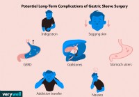
 Žmonėms, sergantiems sunkesnėmis GERL, maistas iš skrandžio gali ištekėti į stemplę arba burną, ypač kai veikla padidina spaudimą pilve, pavyzdžiui, kosint ir pasilenkus.
Su rijimu susiję disfagijos simptomai
Dažniausias rijimo simptomas, atsirandantis dėl disfagijos, yra pojūtis, kad nurytas maistas prilimpa apatinėje kaklo dalyje arba krūtinėje.
Esant neurologinėms problemoms, gali būti sunku pradėti nuryti, nes maistas negali būti varomas liežuviu į gerklę.
Pagyvenę asmenys, turintys protezus, gali blogai sukramtyti maistą ir dėl to praryti didelius įstrigusius kieto maisto gabalėlius.
Disfagija yra medicininis terminas, apibūdinantis rijimo sunkumo simptomą, kilęs iš lotynų ir graikų kalbos žodžių, reiškiančių valgymo sunkumą.
Rijimas yra sudėtingas veiksmas.
Atsižvelgiant į jo sudėtingumą, nenuostabu, kad rijimas, pradedant viršutinės ryklės susitraukimu, buvo „automatizuotas“, o tai reiškia, kad pradėjus rijimą nereikia galvoti apie rijimą. Rijimą kontroliuoja automatiniai refleksai, apimantys nervus ryklėje ir stemplėje, taip pat rijimo centrą smegenyse, kuris nervais yra sujungtas su rykle ir stemple. (Refleksas yra mechanizmas, naudojamas daugeliui organų valdyti. Refleksams reikalingi nervai, esantys organe, pvz., stemplėje, kad pajustų, kas tame organe vyksta, ir siųstų informaciją kitiems nervams, esantiems organo sienelėje arba už jo ribų. Informacija apdorojama šiuose kituose nervuose ir nustatomos atitinkamos reakcijos į organo būklę. Tada dar kiti nervai siunčia pranešimus iš apdorojančių nervų atgal į organą, kad galėtų kontroliuoti organo funkciją, pavyzdžiui, susitraukimą. organo raumenys. Rijimo atveju refleksai pirmiausia apdorojami nervuose, esančiuose ryklės ir stemplės sienelėje, taip pat smegenyse.)
Rijimo sudėtingumas taip pat paaiškina, kodėl yra tiek daug disfagijos priežasčių. Gali kilti problemų dėl:
Problemos gali kilti ryklėje ar stemplėje, pavyzdžiui, dėl fizinio ryklės ar stemplės susiaurėjimo. Disfagija taip pat gali atsirasti dėl raumenų ar nervų, kontroliuojančių ryklės ir stemplės raumenis, ligų arba dėl rijimo centro pažeidimo smegenyse. Galiausiai, ryklėje ir viršutiniame stemplės trečdalyje yra raumenų, kurie yra tokie patys kaip raumenys, kuriuos naudojame savanoriškai (pvz., rankų raumenys), vadinami griaučių raumenimis. Apatinius du trečdalius stemplės sudaro kitokio tipo raumenys, vadinami lygiaisiais raumenimis. Taigi ligos, kurios pirmiausia pažeidžia skeleto raumenis arba lygiuosius kūno raumenis, gali paveikti ryklę ir stemplę, papildydamos disfagijos priežastis.
Yra du simptomai, kurie dažnai laikomi rijimo problemomis (disfagija), kurios tikriausiai nėra. Šie simptomai yra odinofagija ir gaubtelio pojūtis.
Odinofagija reiškia skausmingą rijimą. Kartais žmonėms nelengva atskirti odinofagiją nuo disfagijos. Pavyzdžiui, maistas, kuris prilimpa stemplėje, dažnai yra skausmingas. Ar tai disfagija ar odinofagija, ar abu? Techniškai tai yra disfagija, tačiau žmonės gali tai apibūdinti kaip skausmingą rijimą (t. y. odinofagija). Be to, pacientai, sergantys gastroezofaginio refliukso liga (GERL), gali apibūdinti disfagiją, kai jie iš tikrųjų yra odinofagija. Skausmas, kurį jie jaučia nurijus, išnyksta, kai GERL uždegimas yra gydomas, ir išnyksta, tikriausiai dėl skausmo, kurį sukelia maistas, patenkantis per uždegiminę stemplės dalį.
Odinofagija taip pat gali pasireikšti esant kitoms ligoms, susijusioms su stemplės uždegimu, pavyzdžiui, virusinėmis ir grybelinėmis infekcijomis. Svarbu atskirti disfagiją ir odinofagiją, nes kiekvieno iš jų priežastys gali būti gana skirtingos.
Globus pojūtis reiškia pojūtį, kad gerklėje yra gumbas. Gumbas gali būti nuolat arba tik nurijus. Globuso pojūčio priežastys yra įvairios ir dažnai priežasties nerandama. Globuso pojūtis įvairiai priskiriamas nenormalioms ryklės nervų ar raumenų funkcijoms ir GERL. Globus pojūtis paprastai yra aiškiai apibūdinamas asmenų ir retai sukelia painiavą su tikra disfagija.
Kaip minėta anksčiau, yra daug disfagijos priežasčių. Kad būtų patogiau, disfagijos priežastis galima suskirstyti į dvi grupes;
Priežastys taip pat gali būti skirtingai suskirstytos į kelias grupes.
Esant neurologinėms problemoms, gali būti sunku pradėti nuryti, nes boliuso negalima nustumti liežuviu į gerklę. Pagyvenę asmenys, turintys protezus, gali blogai sukramtyti maistą ir dėl to praryti didelius įstrigusius kieto maisto gabalėlius. (Vis dėlto tai dažniausiai atsitinka, kai yra papildomų problemų ryklėje ar stemplėje, pvz., susiaurėjimas.)
Tačiau dažniausias rijimo simptomas, pasireiškiantis rijimo sutrikimu, yra pojūtis, kad nurytas maistas prilimpa apatinėje kaklo dalyje arba krūtinėje. Jei maistas įstringa gerklėje, gali prasidėti kosulys ar užspringimas, atsikosėjus nurytas maistas. Jei maistas pateks į gerklas, bus išprovokuotas stipresnis kosulys ir užspringimas. Jei minkštasis gomurys neveikia ir tinkamai neužsandarina nosies takų, nurijus maistas, ypač skysčiai, gali patekti į nosį. Kartais nurijus maistas gali vėl patekti į burną iš karto.
Maistas, įstrigęs stemplėje, gali likti ten ilgą laiką. Tai gali sukelti krūtinės prisipildymo pojūtį, kai suvalgoma daugiau maisto, todėl asmuo turi nustoti valgyti ir galbūt gerti skysčius, bandydamas nuplauti maistą. Nesugebėjimas suvalgyti didesnio maisto kiekio gali numesti svorio. Be to, stemplėje likęs maistas naktį gali ištekėti iš stemplės, kai žmogus miega, o žmogus gali būti pažadintas kosėdamas ar užspringęs vidury nakties, kurį išprovokuoja atpylimo maistas. Jei maistas patenka į gerklas, trachėją ir (arba) plaučius, jis gali išprovokuoti astmos epizodus ir netgi sukelti plaučių infekciją bei aspiracinę pneumoniją. Pasikartojanti pneumonija gali sukelti rimtą, nuolatinį ir progresuojantį plaučių pažeidimą. Kartais žmonės nepažadinami iš miego dėl atpylusio maisto, o pabunda ryte, kad ant pagalvės rastų atpylusio maisto.
Asmenys, sulaikantys maistą stemplėje, gali skųstis į rėmenį panašiais (GERL) simptomais. Jų simptomai iš tiesų gali būti dėl GERL, bet labiau tikėtina, kad jie atsiranda dėl pasiliekančio maisto ir netinkamai reaguoja į GERL gydymą.
Esant spazminiams judrumo sutrikimams, asmenims gali pasireikšti krūtinės skausmo epizodai, kurie gali būti tokie stiprūs, kad imituoja širdies priepuolį ir dėl to asmenys turi kreiptis į greitosios pagalbos skyrių. Skausmo dėl spazminių stemplės sutrikimų priežastis neaiški, nors pagrindinė teorija teigia, kad tai yra dėl stemplės raumenų spazmų.
Odinofagija ir globos pojūtis. Jau buvo aptarta, kad kartais sunku atskirti disfagiją nuo odinofagijos, taip pat apie skirtumą tarp disfagijos ir kamuolinio pojūčio.
Trachėjos ir stemplės fistulė. Vienas sutrikimas, kurį galima supainioti su disfagija, yra trachėjos ir stemplės fistulė. Trachėjos ir stemplės fistulė yra atviras ryšys tarp stemplės ir trachėjos, kuris dažnai išsivysto dėl stemplės vėžio, bet gali atsirasti ir kaip įgimtas (įgimtas) apsigimimas. Prarijus maistas gali išprovokuoti kosulį imituojantį kosulį dėl ryklės raumenų disfunkcijos, dėl kurios maistas patenka į gerklas; tačiau fistulės atveju kosulys kyla dėl to, kad maistas iš stemplės per fistulę patenka į trachėją.
Atrajojimo sindromas. Atrajojimo sindromas yra sindromas, kai pavalgius maistas be pastangų grįžta atgal į burną. Paprastai tai pasireiškia jaunesnėms moterims ir gali būti painiojama su disfagija. Tačiau nejaučiama, kad maistas priliptų nurijus.
Gastroezofaginio refliukso liga (GERL). Žmonėms, sergantiems sunkesnėmis GERL, maistas iš skrandžio gali ištekėti į stemplę ar burną, ypač kai veikla padidina spaudimą pilve, pavyzdžiui, kosint ir pasilenkus. Regurgitacija taip pat gali pasireikšti naktį, kai GERL sergantys asmenys miega, kaip ir rijimo sutrikimų turintys asmenys, kurių stemplėje kaupiasi maistas.
Širdies liga. Spaziniai judrumo sutrikimai, sukeliantys disfagiją, gali būti susiję su spontanišku krūtinės skausmu, ty krūtinės skausmu, nesusijusiu su rijimu. Nepaisant disfagijos, spontaniškas krūtinės skausmas visada turi būti laikomas širdies ligos priežastimi, kol nebus pašalinta širdies liga kaip krūtinės skausmo priežastis. Todėl svarbu atidžiai pasitikrinti dėl širdies ligų, prieš laikant stemplę krūtinės skausmo priežastimi, kai disfagija sergantis pacientas skundžiasi spontaniško krūtinės skausmo epizodais.
Disfagija sergančio asmens istorija dažnai suteikia svarbių užuominų apie pagrindinę disfagijos priežastį.
Simptomo ar simptomų pobūdis pateikia svarbiausius užuominas apie disfagijos priežastį. Rijimas, kurį sunku pradėti arba sukelia nosies regurgitaciją, kosulį ar užspringimą, greičiausiai atsiranda dėl burnos ar ryklės problemų. Nurijus, dėl kurio jaučiamas maisto įstrigimas krūtinėje (stemplėje), greičiausiai atsiranda dėl stemplės problemos.
Disfagija, kuri greitai progresuoja per savaites ar kelis mėnesius, rodo piktybinį naviką. Vien tik kieto maisto disfagija rodo fizinį maisto judėjimo sutrikimą, o kieto ir skysto maisto disfagija dažniau kyla dėl stemplės lygiųjų raumenų ligos. Protarpinius simptomus taip pat dažniau sukelia lygiųjų raumenų ligos, o ne stemplės obstrukcija, nes raumenų disfunkcija dažnai būna protarpinė.
Jau esamos ligos taip pat suteikia užuominų. Sergantiesiems skeleto raumenų ligomis (pavyzdžiui, polimiozitu), smegenų (dažniausiai insultu) ar nervų sistemos ligomis dažniau pasireiškia disfagija dėl burnos ir ryklės raumenų ir nervų disfunkcijos. Žmonės, sergantys kolageno kraujagyslių ligomis, pavyzdžiui, sklerodermija, dažniau turi problemų su stemplės raumenimis, ypač neveiksminga peristaltika.
Pacientams, kuriems anksčiau buvo GERL, dažniau pasireiškia stemplės susiaurėjimas kaip disfagijos priežastis, nors apie 20 % pacientų, kuriems yra striktūros, GERL simptomai pasireiškia minimaliai arba jų visai nėra prieš prasidedant disfagijai. Manoma, kad refliuksas, atsirandantis naktį, labiau pažeidžia stemplę. Taip pat yra didesnė rizika susirgti stemplės vėžiu asmenims, sergantiems ilgalaike GERL.
Svorio netekimas gali būti sunkios disfagijos arba piktybinio naviko požymis. Dažniau nei numesti svorio, žmonės aprašo savo valgymo įpročių pasikeitimą – mažesni kąsniai, papildomas kramtymas – dėl kurių valgymas pailgėja taip, kad prie stalo jie baigia valgyti paskutiniai. Pastarasis modelis, jei jis pasireiškia ilgą laiką, rodo nepiktybinę, gana stabilią arba lėtai progresuojančią disfagijos priežastį. Krūtinės skausmo epizodai, atsirandantys ne dėl širdies ligų, rodo stemplės raumenų ligas. Gimimas ir apsigyvenimas Centrinėje ar Pietų Amerikoje yra susijęs su Chagas liga.
Fizinis patikrinimas yra ribotas, kad būtų galima nustatyti disfagijos priežastis. Neurologinio tyrimo anomalijos rodo neurologines ar raumenų ligas. Stebint individualų rijimą, galima nustatyti, ar sunku pradėti rijimą, tai yra neurologinės ligos požymis. Kaklo navikai rodo ryklės suspaudimo galimybę. Trachėja, kurios negalima perkelti iš vienos pusės į kitą ranka, rodo, kad krūtinės ląstos apačioje yra auglys, įstrigęs trachėja ir galbūt stemplė. Stebint atrofiją (sumažėjusį dydį) arba liežuvio susižavėjimą (smulkus drebulys) taip pat galima spręsti apie nervų sistemos arba skeleto raumenų ligas.
Endoskopija. Endoskopijos metu per burną, ryklę, stemplę ir į skrandį įkišamas ilgas (vieno metro), lankstus vamzdelis su šviesa ir kamera jo gale. Ryklės ir stemplės gleivinę galima įvertinti vizualiai, galima paimti biopsijas (mažus audinio gabalėlius), kad būtų galima ištirti mikroskopu arba bakterijų ar virusų kultūras.
Endoskopija yra puiki priemonė diagnozuoti navikus, susiaurėjimus ir Schatzki žiedus bei stemplės infekcijas. Tai taip pat labai tinka vidurinės ir apatinės stemplės divertikulams diagnozuoti, tačiau blogai diagnozuojant viršutinės stemplės divertikulus (Zenkerio divertikulą).
Galima stebėti stemplės raumenų susitraukimo anomalijas, tačiau stemplės manometrija yra testas, kuris daug geriau tinka stemplės raumenų funkcijai įvertinti. Resistance passing the endoscope through the lower esophageal sphincter combined with a lack of esophageal contractions is a fairly reliable sign of achalasia or Chagas disease (due to the inability of the lower esophageal sphincter to relax), but it is important when there is resistance to exclude the presence of a stricture or cancer which also can cause resistance. Finally, there is a characteristic appearance of the esophageal lining when infiltrated with eosinophils that strongly suggests the presence of eosinophilic esophagitis.
X-rays. There are two different types of X-rays that can be done to diagnose the cause of dysphagia. The barium swallow or esophagram is the simplest type. For the barium swallow, mouthfuls of barium are swallowed, and X-ray films are taken of the esophagus at several points in time while the bolus of barium traverses the esophagus. The barium swallow is excellent for diagnosing moderate-to-severe external compression, tumors, and strictures of the esophagus. Occasionally, however, Schatzki's rings can be missed.
Another type of X-ray study that can be done to evaluate swallowing is the video esophagram or video swallow, sometimes called a video-fluoroscopic swallowing study. For the video swallow, instead of several static X-ray images of the bolus traversing the esophagus, a video X-ray is taken. The video study can be reviewed frame by frame and is able to show much more than the barium swallow. This usually is not important for diagnosing tumors or strictures, which are well seen on barium swallow, but it is more effective for suggesting problems with the contraction of the muscles of the esophagus and pharynx (though esophageal manometry, discussed later, is still better for studying contraction), milder external compression of the esophagus, and Schatzki's rings. The video study can be extended to include the pharynx where it is the best method for demonstrating osteophytes, cricopharyngeal bars, and Zenker's diverticuli. A modified barium swallow is a version of the test evaluating the oropharyngeal phases of swallowing. A speech pathologist is usually involved with the evaluation to determine subtle sequence and phase abnormalities.
The video swallow also is excellent for diagnosing penetration of barium (the equivalent of food) into the larynx and trachea due to neurological and muscular problems of the pharynx that may be causing coughing or choking after swallowing food.
Esophageal manometry. Esophageal manometry, also known as esophageal motility testing, is a means to evaluate the function of pharyngeal and esophageal muscles. For manometry, a thin, flexible catheter is passed through the nose and pharynx and into the esophagus. The catheter is able to sense pressure at multiple locations along its length in both the pharynx and the esophagus. When the pharyngeal and esophageal muscles contract, they generate a pressure on the catheter which is sensed, measured and recorded from each location. The magnitude of the pressure at each pressure-sensing location and the timing of the increases in pressure at each location in relation to other locations give an accurate picture of how the muscles of the pharynx and esophagus are contracting.
The value of manometry is in diagnosing and differentiating among diseases of the muscle or the nerves controlling the muscles that result in muscle dysfunction of the pharynx and esophagus. Thus, it is useful for diagnosing the swallowing dysfunction caused by diseases of the brain, skeletal muscle of the pharynx, and smooth muscle of the esophagus.
Esophageal impedence. Esophageal impedence testing utilizes catheters similar to those used for esophageal manometry. Impedence testing, however, senses the flow of the bolus through the esophagus. Thus, it is possible to determine how well the bolus is traversing the esophagus and correlate the movement with concomitantly recorded esophageal pressures determined by manometry. (It also can be used to sense reflux of stomach contents into the esophagus among patients with GERD.) Multiple sites along the length of the esophagus can be tested to assess the movement of the bolus and presence of reflux, including how high up it extends.
Esophageal acid testing. Esophageal acid testing is not a test that directly diagnoses diseases of the esophagus. Rather, it is a method for determining whether or not there is reflux of acid from the stomach into the esophagus, a cause of the most common esophageal problem leading to dysphagia, esophageal stricture. For acid testing, a thin catheter is inserted through the nose, down the throat, and into the esophagus. At the tip of the catheter and placed just above the junction of the esophagus with the stomach is an acid-sensing probe. The catheter coming out of the nose passes back over the ear and down to the waist where it is attached to a recorder. Each time acid refluxes (regurgitates) from the stomach and into the esophagus it hits the probe, and the reflux of acid is recorded by the recorder. At the end of a prolonged period, usually 24 hours, the catheter is removed and the information from the recorder is downloaded into a computer for analysis. Most people have a small amount of reflux of acid, but individuals with GERD have more. Thus, acid testing can determine if GERD is likely to be the cause of the esophageal problem such as a stricture, as well as if treatment of GERD is adequate by showing the amount of acid that refluxes during treatment is normal.
An alternative method of esophageal acid testing uses a small capsule containing an acid-sensing probe that is attached to the esophageal lining just above the junction of the esophagus with the stomach. The capsule wirelessly transmits the presence of episodes of acid regurgitation to a receiver carried on the chest. The capsule records for two or three days and later is shed into the esophagus and passes out of the body in the stool.
Other tests The diagnosis of muscular dystrophies and metabolic myopathies usually involves a combination of tests including blood tests that can suggest muscle injury, electromyograms to determine if nerves and muscles are working normally, biopsies of muscles, and genetic testing.
The treatment of dysphagia varies and depends on the cause of the dysphagia. One option for supporting patients either transiently or long-term until the cause of the dysphagia resolves is a feeding tube. The tube for feeding may be passed nasally into the stomach or through the abdominal wall into the stomach or small intestine. Once oral feeding resumes, the tube can be removed.
Treatment for obstruction of the pharynx or esophagus requires removal of the obstruction.
Tumors usually are removed surgically although occasionally they can be removed endoscopically, totally or partially. Radiation therapy and chemotherapy also may be used particularly for malignant tumors of the pharynx and its surrounding tissues. If malignant tumors of the esophagus cannot be easily removed or the tumor has spread and survival will be limited, swallowing can be improved by placing stents within the esophagus across the area of obstruction. Occasionally, obstructing tumors can be dilated the same way as strictures. (See below.)
Strictures and Schatzki's rings usually are treated with endoscopic dilation, a procedure in which the narrowed area is stretched either by a long, semi-rigid tube passed through the mouth or a balloon that is blown up inside the esophagus.
The most common infiltrating disease causing dysphagia is eosinophilic esophagitis which usually is successfully treated with swallowed corticosteroids. The role of food allergy as a cause of eosinophilic esophagitis is debated; however, there are reports of using elimination diets to identify specific foods that are associated with allergy. Elimination of these foods has been reported to prevent or reverse the infiltration of the esophagus with eosinophils, particularly in children.
Diverticuli of the pharynx and esophagus usually are treated surgically by excising them. Occasionally they can be treated endoscopically. Cricopharyngeal bars are treated surgically by cutting the thickened muscle. Osteophytes also can be removed surgically.
Congenital abnormalities of the esophagus usually are treated surgically soon after birth so that oral feeding can resume.
As previously discussed, strokes are the most common disease of the brain to cause dysphagia. Dysphagia usually is at its worst immediately after the stroke, and often the dysphagia improves with time and even may disappear. If it does not disappear, swallowing is evaluated, usually with a video swallowing study. The exact abnormality of function can be defined and different maneuvers can be performed to see if they can counter the effects of the dysfunction. For example, in some patients it is possible to prevent aspiration of food by turning the head to the side when swallowing or by drinking thickened liquids (since thin liquids is the food most likely to be aspirated).
Tumors of the brain, in some cases, can be removed surgically; however, it is unlikely that surgery will reverse the dysphagia. Parkinson's disease and multiple sclerosis can be treated with drugs and may be useful in patients with dysphagia.
Achalasia is treated like a stricture of the esophagus with dilation, usually with a balloon. A second option is surgical treatment in which the muscle of the lower esophageal sphincter is cut (a myotomy) in order to reduce the pressure and obstruction caused by the non-relaxing sphincter. Drugs that relax the sphincter usually have little or a transient effect and are useful only when achalasia is mild.
An option for individuals who are at high risk for surgery or balloon dilation is injection of botulinin toxin into the sphincter. The toxin paralyzes the muscle of the sphincter and causes the pressure within the sphincter to decrease. The effects of botulinin toxin are transient, however, and repeated injections usually are necessary. It is best to treat achalasia early before the obstruction causes the esophagus to enlarge (dilate) which can lead to additional problems such as food collecting above the sphincter with regurgitation and aspiration.
In other spastic motility disorders, several drugs may be tried, including anti-cholinergic medications, peppermint, nitroglycerin, and calcium channel blockers, but the effectiveness of these drugs is not clear and studies with them are nonexistent or limited.
For patients with severe and uncontrollable symptoms of pain and/or dysphagia, a surgical procedure called a long myotomy occasionally is performed. A long myotomy is similar to the surgical treatment for achalasia but the cut in the muscle is extended up along the body of the esophagus for a variable distance in an attempt to reduce pressures and obstruction to the bolus.
There is no treatment for ineffective peristalsis, and individuals must change their eating habits. Fortunately, ineffective peristalsis infrequently causes severe dysphagia by itself. When moderate or severe dysphagia is associated with ineffective peristalsis it is important to be certain that there is no additional obstruction of the esophagus, for example, by a stricture due to GERD, that is adding to the effects of reduced muscle function and making dysphagia worse than the ineffective peristalsis alone. Most causes of obstruction can be treated.
There are effective drug therapies for polymyositis and myasthenia gravis that should also improve associated dysphagia. Treatment of the muscular dystrophies is primarily directed at preventing deformities of the joints that commonly occur and lead to immobility, but there are no therapies that affect the dysphagia. Corticosteroids and drugs that suppress immunity sometimes are used to treat some of the muscular dystrophies, but their effectiveness has not been demonstrated.
There is no treatment for the metabolic myopathies other than changes in lifestyle and diet.
Diseases that reduce the production of saliva can be treated with artificial saliva or over-the-counter and prescription drugs that stimulate the production of saliva.
There is no treatment for Alzheimer's disease.
With the exception of dysphagia caused by stroke for which there can be marked improvement, dysphagia from other causes is stable or progressive, and the prognosis depends on the underlying cause, its tendency to progress, the availability of therapy, and the response to therapy.
Recent developments in the diagnostic arena are beginning to bring new insights into esophageal function, specifically, high resolution and 3D manometry, and endoscopic ultrasound.
High resolution and 3D manometry
High resolution and 3D manometry are extensions of standard manometry that utilize similar catheters. The difference is that the pressure-sensing locations on the catheters are very close together and ring the catheter. Recording of pressures from so many locations gives an extremely detailed picture of how esophageal muscle is contracting. The primary value of these diagnostic procedures is that they "integrate" the activities of the esophagus so that the overall pattern of swallowing can be recognized, which is particularly important in complex motility disorders. In addition, their added detail allows the recognition of subtle abnormalities and hopefully will be able to help define the clinical importance of subtle abnormalities of muscle contraction associated with lesser degrees of dysphagia.
Endoscopic ultrasonography has been available for many years but has recently been applied to the evaluation of esophageal muscle diseases. Ultrasound uses sound waves to penetrate tissues. The sound waves are reflected by the tissues and structures they encounter, and, when analyzed, the reflections give information about the tissues and structures from which they are reflected. In the esophagus, endoscopic ultrasonography has been used to determine the extent of penetration of tumors into the esophageal wall and the presence of metastases to adjacent lymph nodes. More recently, endoscopic ultrasonography has been used to obtain a detailed look at the muscles of the esophagus. What has been found is that in some disorders, particularly the spastic motility disorders, the muscle of the esophagus is thickened. Moreover, thickening of the muscle sometimes can be recognized only by ultrasonography even when spastic abnormalities are not seen with manometry. The exact role of endoscopic ultrasonography has not yet been determined but is an exciting area for future research.
 Ar saldūs gėrimai kenkia mūsų storajai žarnai?
 Geriausi JAV GI gydytojai / geriausiai įvertinti gastroenterologai – dr. Vikramas Tarugu
 Ilgalaikės komplikacijos po skrandžio rankovės operacijos
 Klimato kaita, žygiai pėsčiomis Pavojus dėl mėsą valgančių bakterijų infekcijų
 Kai kurios bakterijų rūšys gali padidinti ŽIV riziką moterims,
 Kaip RD naudojo maistą, kad užbaigtų 25 metus trukusį vidurių užkietėjimą
 Skelbiama:Chriso Kresserio sveikatos radijo laida „Revolution“
„Neįsižeisk, daktaras, bet atrodo, kad nesupranti, kas su manimi negerai“. Tai aš sakiau, kai 2008 m. atleidau savo gydytoją... Vaikystėje buvau išmokytas pasitikėti savo gydytoju... Jausčiausi sau
 Ar mūsų imuninė sistema atlieka svarbų vaidmenį sergant IBS?
IBS yra sudėtingas, nevienalytis sutrikimas, reiškiantis, kad ne kiekvienas atvejis atrodo vienodai. Vykstant tyrimams suprantame, kad IBS diagnozės greičiausiai bus suskirstytos į potipius, priklauso
 Šlapimo takų infekcijos (UTI) vaikams
102,2 F arba 39 C) ir pilvo skausmas. Šlapimo takų infekcijos yra gana dažna problema vaikystėje ir gali turėti gerybinę eigą, reaguoti į paprastą gydymą antibiotikais, arba būti susijusios su reik