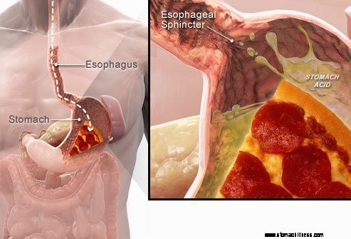
 Когда вы глотаете пищу, она проходит по пищеводу и проходит через мышечное кольцо, известное как нижний пищеводный сфинктер ( ЛЕС). Эта структура открывается, чтобы позволить пище пройти в желудок. Он должен оставаться закрытым, чтобы содержимое желудка оставалось там, где ему и место.
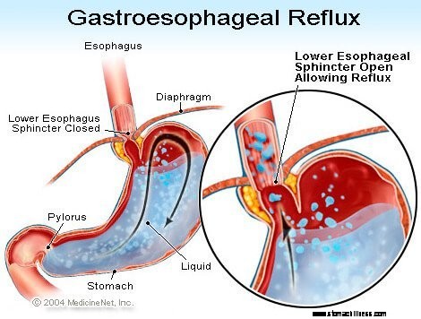
Симптомы ГЭРБ или кислотного рефлюкса вызваны регургитацией кислого жидкого содержимого желудка обратно в пищевод. Наиболее распространенным симптомом ГЭРБ является изжога.
Другие симптомы, которые могут возникнуть в результате ГЭРБ, включают:
 Наиболее частым симптомом ГЭРБ является изжога.
 Гастроэзофагеальная рефлюксная болезнь, обычно называемая ГЭРБ или кислотным рефлюксом, представляет собой состояние, при котором жидкое содержимое желудка регургитирует (обратно или рефлюксно) в пищевод. Жидкость может воспаляться и повреждать слизистую (эзофагит), хотя видимые признаки воспаления встречаются у меньшинства пациентов. Срыгиваемая жидкость обычно содержит кислоту и пепсин, которые вырабатываются желудком. (Пепсин — это фермент, который начинает переваривать белки в желудке.) Рефлюксная жидкость также может содержать желчь, которая попала в желудок из двенадцатиперстной кишки. Первая часть тонкой кишки прикрепляется к желудку. Кислота считается наиболее вредным компонентом рефлюксной жидкости. Пепсин и желчь также могут повредить пищевод, но их роль в воспалении и повреждении пищевода не так очевидна, как роль кислоты.
ГЭРБ является хроническим заболеванием. Как только это начинается, это, как правило, на всю жизнь. Если имеется повреждение слизистой оболочки пищевода (эзофагит), это также является хроническим заболеванием. Более того, после того, как пищевод зажил с помощью лечения и лечение было прекращено, у большинства пациентов травма возвращается в течение нескольких месяцев. После начала лечения ГЭРБ его необходимо продолжать в течение неопределенного времени. Однако некоторых пациентов с интермиттирующими симптомами и отсутствием эзофагита можно лечить только в течение симптоматических периодов.
Фактически рефлюкс жидкого содержимого желудка в пищевод происходит у большинства нормальных людей. Одно исследование показало, что рефлюкс часто возникает как у здоровых людей, так и у пациентов с ГЭРБ. Однако у больных ГЭРБ рефлюксная жидкость чаще содержит кислоту, и кислота дольше остается в пищеводе. Также было обнаружено, что у пациентов с ГЭРБ уровень рефлюкса жидкости в пищевод выше, чем у здоровых людей.
Как это часто бывает, у организма есть способы защитить себя от вредного воздействия рефлюкса и кислоты. Например, чаще всего рефлюкс возникает в течение дня, когда люди находятся в вертикальном положении. В вертикальном положении рефлюксная жидкость с большей вероятностью стекает обратно в желудок из-за действия силы тяжести. Кроме того, в то время как люди бодрствуют, они неоднократно сглатывают, независимо от того, есть рефлюкс или нет. Каждый глоток переносит рефлюксную жидкость обратно в желудок. Наконец, слюнные железы во рту производят слюну, содержащую бикарбонат. С каждым глотком слюна, содержащая бикарбонат, проходит по пищеводу. Бикарбонат нейтрализует небольшое количество кислоты, которая остается в пищеводе после того, как большая часть кислой жидкости удалена под действием силы тяжести и проглатывания.
Гравитация, глотание и слюна являются важными защитными механизмами для пищевода, но они эффективны только тогда, когда человек находится в вертикальном положении. Ночью во время сна сила тяжести не действует, глотание прекращается, а секреция слюны снижается. Следовательно, рефлюкс, возникающий ночью, с большей вероятностью приведет к тому, что кислота останется в пищеводе дольше и вызовет большее повреждение пищевода.
Определенные условия делают человека восприимчивым к ГЭРБ. Например, ГЭРБ может быть серьезной проблемой во время беременности. Повышенный уровень гормонов при беременности, вероятно, вызывает рефлюкс за счет снижения давления в нижнем пищеводном сфинктере (см. ниже). В то же время растущий плод увеличивает давление в брюшной полости. Ожидается, что оба этих эффекта будут усиливать рефлюкс. Также более склонны к развитию ГЭРБ пациенты с заболеваниями, ослабляющими мышцы пищевода, такими как склеродермия или смешанные заболевания соединительной ткани.
Причина ГЭРБ сложна и может включать несколько причин. Более того, разные причины могут воздействовать на разных людей или даже на одного и того же человека в разное время. У небольшого числа пациентов с ГЭРБ вырабатывается аномально большое количество кислоты, но это необычно и не является сопутствующим фактором у подавляющего большинства пациентов.
Факторы, способствующие ГЭРБ:
Действие нижнего пищеводного сфинктера (НПС), возможно, является наиболее важным фактором (механизмом) предотвращения рефлюкса. Пищевод представляет собой мышечную трубку, которая тянется от нижнего отдела глотки до желудка. НПС представляет собой специализированное кольцо мышц, которое окружает самый нижний конец пищевода в месте его соединения с желудком. Мышца, входящая в состав НПС, активна большую часть времени, то есть в состоянии покоя. Это означает, что он сокращается и перекрывает проход из пищевода в желудок. Это закрытие прохода предотвращает рефлюкс. При проглатывании пищи или слюны НПС расслабляется на несколько секунд, позволяя пище или слюне пройти из пищевода в желудок, а затем снова закрывается.
У пациентов с ГЭРБ было обнаружено несколько различных аномалий НПС. Два из них связаны с функцией НПС. Во-первых, это аномально слабое сокращение НПС, что снижает его способность предотвращать рефлюкс. Во-вторых, это аномальные релаксации НПС, называемые транзиторными релаксациями НПС. Они аномальны тем, что не сопровождают ласточки и длятся долго, до нескольких минут. Эти длительные релаксации способствуют более легкому рефлюксу. Преходящие релаксации НПС возникают у больных ГЭРБ чаще всего после еды, когда желудок растянут пищей. Временное расслабление НПС также встречается у людей без ГЭРБ, но нечасто.
Наиболее недавно описанной аномалией у пациентов с ГЭРБ является вялость НПС. В частности, одинаковое растягивающее давление открывает НПС больше у пациентов с ГЭРБ, чем у лиц без ГЭРБ. По крайней мере, теоретически это облегчило бы открытие НПС и/или увеличило обратный поток кислоты в пищевод, когда НПС открыт.
Грыжи пищеводного отверстия диафрагмы способствуют рефлюксу, хотя то, каким образом они способствуют, неясно. У большинства пациентов с ГЭРБ есть грыжи пищеводного отверстия диафрагмы, но у многих их нет. Следовательно, не обязательно иметь грыжу пищеводного отверстия диафрагмы, чтобы иметь ГЭРБ. Более того, у многих людей есть грыжа пищеводного отверстия диафрагмы, но нет ГЭРБ. Точно неизвестно, как и почему развиваются грыжи пищеводного отверстия диафрагмы.
В норме НПС располагается на том же уровне, где пищевод проходит из грудной клетки через небольшое отверстие в диафрагме в брюшную полость. (Диафрагма представляет собой мышечную горизонтальную перегородку, отделяющую грудную клетку от брюшной полости.) При грыже пищеводного отверстия небольшая часть верхней части желудка, которая прикрепляется к пищеводу, проталкивается вверх через диафрагму. В результате небольшая часть желудка и НПС оказываются в грудной клетке, а НПС больше не находится на уровне диафрагмы.
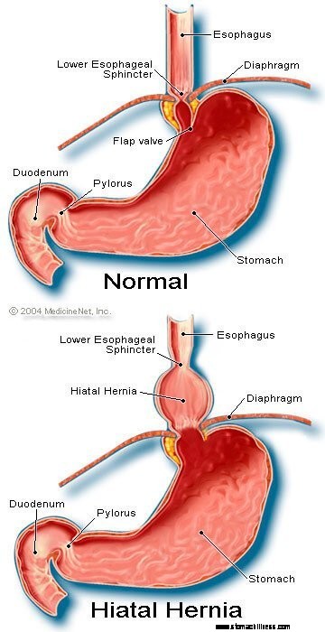 Изображение грыжи пищеводного отверстия диафрагмы
Похоже, что диафрагма, окружающая НПС, играет важную роль в предотвращении рефлюкса. То есть у людей без грыж пищеводного отверстия диафрагма, окружающая пищевод, постоянно сокращается, но затем расслабляется при глотании, как и НПС. Обратите внимание, что эффекты НПС и диафрагмы проявляются в одном и том же месте у пациентов без грыж пищеводного отверстия диафрагмы. Следовательно, барьер рефлюкса равен сумме давлений, создаваемых НПС и диафрагмой. Когда НПС перемещается в грудную клетку с грыжей пищеводного отверстия диафрагмы, диафрагма и НПС продолжают оказывать давление и барьерный эффект. Однако теперь они делают это в разных местах. Следовательно, давления больше не являются аддитивными. Вместо этого один барьер высокого давления для рефлюкса заменяется двумя барьерами более низкого давления, и, таким образом, рефлюкс происходит легче. Таким образом, снижение барьера давления — это один из способов, которым грыжа пищеводного отверстия диафрагмы может способствовать рефлюксу.
Как упоминалось ранее, глотание играет важную роль в выведении кислоты из пищевода. Глотание вызывает кольцевидную волну сокращения мышц пищевода, что сужает просвет (внутреннюю полость) пищевода. Сокращение, называемое перистальтикой, начинается в верхних отделах пищевода и распространяется на нижние отделы пищевода. Он проталкивает пищу, слюну и все, что находится в пищеводе, в желудок.
Когда волна сокращения нарушена, рефлюксная кислота не выталкивается обратно в желудок. У пациентов с ГЭРБ описано несколько нарушений сокращения. Например, волны сокращения могут не начинаться после каждого глотка или волны сокращения могут затухать, не достигнув желудка. Кроме того, давление, создаваемое сокращениями, может быть слишком слабым, чтобы вытолкнуть кислоту обратно в желудок. Такие нарушения сокращения, которые уменьшают клиренс кислоты из пищевода, часто обнаруживаются у пациентов с ГЭРБ. На самом деле они чаще всего обнаруживаются у пациентов с наиболее тяжелой формой ГЭРБ. Ожидается, что последствия аномальных сокращений пищевода будут усиливаться ночью, когда гравитация не помогает возвращать рефлюксную кислоту в желудок. Обратите внимание, что курение также существенно снижает клиренс кислоты из пищевода. Этот эффект сохраняется не менее 6 часов после выкуривания последней сигареты.
Чаще всего рефлюкс в течение дня возникает после еды. Этот рефлюкс, вероятно, связан с преходящей релаксацией НПС, вызванной растяжением желудка пищей. Было обнаружено, что у меньшинства пациентов с ГЭРБ желудок опорожняется аномально медленно после еды. Это называется гастропарез. Более медленное опорожнение желудка продлевает наполнение желудка пищей после еды. Следовательно, более медленное опорожнение продлевает период времени, в течение которого более вероятно возникновение рефлюкса. Есть несколько лекарств, связанных с нарушением опорожнения желудка, например:
Люди не должны прекращать прием этих или любых назначенных лекарств, пока лечащий врач не обсудит с ними возможную ситуацию с ГЭРБ.
Симптомы неосложненной ГЭРБ в основном следующие:
Другие симптомы возникают при осложнениях ГЭРБ и будут обсуждаться с осложнениями.
Когда кислота рефлюксирует обратно в пищевод у пациентов с ГЭРБ, нервные волокна в пищеводе стимулируются. Эта нервная стимуляция чаще всего приводит к изжоге, боли, характерной для ГЭРБ. Изжога обычно описывается как жгучая боль в середине грудной клетки. Он может начинаться высоко в животе или может распространяться на шею. Однако у некоторых пациентов боль может быть острой или давящей, а не жгучей. Такая боль может имитировать сердечную боль (стенокардию). У других пациентов боль может отдавать в спину.
Поскольку кислотный рефлюкс чаще возникает после еды, изжога также чаще возникает после еды. Изжога также чаще возникает, когда люди лежат, потому что без воздействия гравитации рефлюкс возникает легче, и кислота возвращается в желудок медленнее. Многие пациенты с ГЭРБ просыпаются от изжоги.
Эпизоды изжоги, как правило, случаются периодически. Это означает, что эпизоды становятся более частыми или тяжелыми в течение нескольких недель или месяцев, а затем становятся менее частыми или тяжелыми или даже отсутствуют в течение нескольких недель или месяцев. Эта периодичность симптомов дает основание для прерывистого лечения у пациентов с ГЭРБ, у которых нет эзофагита. Тем не менее изжога остается проблемой на всю жизнь и почти всегда возвращается.
Срыгивание – это появление рефлюксной жидкости во рту. У большинства пациентов с ГЭРБ обычно лишь небольшое количество жидкости достигает пищевода, и жидкость остается в нижних отделах пищевода. Иногда у некоторых пациентов с ГЭРБ большие количества жидкости, иногда содержащие пищу, рефлюксируют и достигают верхних отделов пищевода.
В верхнем конце пищевода находится верхний пищеводный сфинктер (ВПС). ВПС представляет собой кольцевое мышечное кольцо, действие которого очень похоже на НПС. То есть УЭС предотвращает заброс содержимого пищевода в горло. Когда небольшие количества рефлюксной жидкости и/или пищи проходят через СПС и попадают в горло, во рту может ощущаться кислый привкус. Если большие количества нарушают ЕЭС, пациенты могут внезапно обнаружить, что их рты заполнены жидкостью или пищей. Более того, частое или продолжительное срыгивание может привести к эрозии зубов, вызванной кислотой.
Тошнота при ГЭРБ встречается редко. У некоторых пациентов, однако, это может быть частым или тяжелым и может привести к рвоте. Фактически, у пациентов с необъяснимой тошнотой и/или рвотой ГЭРБ является одним из первых состояний, которые следует учитывать. Неясно, почему у некоторых пациентов с ГЭРБ в основном развивается изжога, а у других — в основном тошнота.
Жидкость из желудка, которая рефлюксирует в пищевод, повреждает клетки, выстилающие пищевод. Организм реагирует так же, как обычно реагирует на повреждение, то есть воспалением (эзофагитом). Цель воспаления – нейтрализовать повреждающий агент и начать процесс заживления. Если повреждение уходит глубоко в пищевод, образуется язва. Язва — это просто разрыв слизистой оболочки пищевода, который возникает в области воспаления. Язвы и вызванное ими дополнительное воспаление могут разрушать кровеносные сосуды пищевода и вызывать кровотечение в пищевод.
Иногда кровотечение сильное и может потребоваться:
Язвы пищевода заживают с образованием рубцов (фиброз). Со временем рубцовая ткань сморщивается и сужает просвет (внутреннюю полость) пищевода. Это рубцовое сужение называется стриктурой. Проглоченная пища может застрять в пищеводе, как только сужение станет достаточно сильным (обычно когда просвет пищевода сужается до одного сантиметра в диаметре). В этой ситуации может потребоваться эндоскопическое удаление застрявшей пищи. Затем, чтобы еда не прилипала, сужение необходимо растянуть (расширить). Кроме того, чтобы предотвратить рецидив стриктуры, также необходимо предотвратить рефлюкс.
Давно существующая и/или тяжелая ГЭРБ вызывает у некоторых пациентов изменения в клетках, выстилающих пищевод. Эти клетки являются предраковыми и могут, хотя обычно, становиться раковыми. Это состояние называется пищеводом Барретта и встречается примерно у 10% пациентов с ГЭРБ. Тип рака пищевода, связанный с пищеводом Барретта (аденокарцинома), увеличивается по частоте. Непонятно, почему у некоторых пациентов с ГЭРБ развивается пищевод Барретта, но у большинства его нет.
Пищевод Барретта можно распознать визуально во время эндоскопии и подтвердить микроскопическим исследованием клеток слизистой оболочки. Затем пациенты с пищеводом Барретта могут подвергаться периодическому наблюдению эндоскопии с биопсией, хотя нет согласия относительно того, какие пациенты нуждаются в наблюдении. Целью наблюдения является выявление прогрессирования предраковых изменений в более злокачественные, чтобы можно было начать противораковое лечение. Также считается, что пациенты с пищеводом Барретта должны получать максимальное лечение ГЭРБ, чтобы предотвратить дальнейшее повреждение пищевода. Изучаются процедуры, которые удаляют аномальные клетки слизистой оболочки. Для удаления клеток можно использовать несколько эндоскопических нехирургических методов. Эти методы привлекательны тем, что не требуют хирургического вмешательства; однако они связаны с осложнениями, а долгосрочная эффективность лечения еще не определена. Хирургическое удаление пищевода всегда возможно.
Многие нервы находятся в нижнем отделе пищевода. Некоторые из этих нервов стимулируются рефлюксной кислотой, и это раздражение вызывает боль (обычно изжогу). Другие нервы, которые стимулируются, не вызывают боли. Вместо этого они стимулируют еще другие нервы, провоцирующие кашель. Таким образом, рефлюксная жидкость может вызывать кашель, даже не достигая горла! Аналогичным образом рефлюкс в нижний отдел пищевода может стимулировать пищеводные нервы, которые соединяются с легкими, и могут стимулировать нервы, идущие к легким. Эти нервы к легким затем могут вызвать сужение меньших дыхательных трубок, что приводит к приступу астмы.
Хотя ГЭРБ может вызывать кашель, это не частая причина необъяснимого кашля. Хотя ГЭРБ также может быть причиной астмы, более вероятно, что она провоцирует астматические приступы у пациентов, уже страдающих астмой. Хотя хронический кашель и астма являются распространенными заболеваниями, неясно, насколько часто они усугубляются или вызываются ГЭРБ.
Если рефлюксная жидкость проходит через верхний пищеводный сфинктер, она может попасть в горло (глотку) и даже в гортань (гортань). Возникающее воспаление может привести к боли в горле и охриплости. Как и в случае с кашлем и астмой, неясно, насколько часто ГЭРБ вызывает необъяснимое воспаление горла и гортани.
Рефлюксная жидкость, которая проходит из горла (глотки) в гортань, может попасть в легкие (аспирация). Рефлюкс жидкости в легкие (называемый аспирацией) часто приводит к кашлю и удушью. Однако аспирация может происходить и без этих симптомов. С этими симптомами или без них аспирация может привести к инфекции легких и пневмонии. Этот тип пневмонии является серьезной проблемой, требующей немедленного лечения. Когда аспирация не сопровождается симптомами, это может привести к медленному прогрессирующему рубцеванию легких (легочный фиброз), которое можно увидеть на рентгенограммах органов грудной клетки. Аспирация чаще происходит ночью, потому что в это время процессы (механизмы), защищающие от рефлюкса, не активны, а кашлевой рефлекс, защищающий легкие, также неактивен.
Горло сообщается с носовыми ходами. У маленьких детей два участка лимфатической ткани, называемые аденоидами, расположены там, где верхняя часть глотки соединяется с носовыми ходами. Ходы из пазух и трубы из среднего уха (евстахиевы трубы) открываются в заднюю часть носовых ходов возле аденоидов. Рефлюксная жидкость, попадающая в верхние отделы глотки, может воспалить аденоиды и вызвать их набухание. Затем набухшие аденоиды могут блокировать проходы из пазух и евстахиевых труб. Когда придаточные пазухи и среднее ухо закрываются от носовых ходов набуханием аденоидов, в них скапливается жидкость. Это скопление жидкости может привести к дискомфорту в носовых пазухах и ушах. Поскольку аденоиды заметны у маленьких детей, а не у взрослых, это скопление жидкости в ушах и носовых пазухах наблюдается у детей, а не у взрослых.
Существует множество процедур, тестов и оценки симптомов (например, изжоги) для диагностики и оценки состояния пациентов с ГЭРБ.
Обычно ГЭРБ проявляется характерным симптомом изжогой. Изжога чаще всего описывается как загрудинное (под серединой груди) жжение, возникающее после еды и часто усиливающееся в положении лежа. Для подтверждения диагноза врачи часто назначают пациентам препараты для подавления выработки кислоты желудком. Если после этого изжога значительно уменьшилась, диагноз ГЭРБ считается подтвержденным. Этот подход к постановке диагноза на основе реакции симптомов на лечение обычно называется терапевтическим испытанием.
У этого подхода есть проблемы. Например, пациенты с состояниями, которые могут имитировать ГЭРБ, в частности язвы двенадцатиперстной кишки или желудка (желудка), также могут реально реагировать на такое лечение. В этой ситуации, если врач предположит, что проблема связана с ГЭРБ, причина язвенной болезни будет упущена, например, тип инфекции, называемый Helicobacter pylori. (H. pylori ), нестероидные противовоспалительные препараты или НПВП (например, ибупрофен), также могут вызывать язвы, и эти состояния будут лечиться иначе, чем ГЭРБ.
Более того, как и при любом лечении, возможно, существует 20-процентный эффект плацебо, а это означает, что 20 % пациентов будут реагировать на плацебо (неактивную) таблетку или вообще на любое лечение. Это означает, что у 20% пациентов, у которых есть другие причины симптомов, кроме ГЭРБ (или язвы), симптомы уменьшатся после лечения ГЭРБ. Таким образом, на основании их реакции на лечение (терапевтическое испытание) эти пациенты затем будут продолжать лечиться от ГЭРБ, даже если у них нет ГЭРБ. Более того, истинная причина их симптомов не будет выяснена.
Эндоскопия верхних отделов желудочно-кишечного тракта (также известная как эзофагогастродуоденоскопия или ЭГДС) является распространенным методом диагностики ГЭРБ. ФГДС — это процедура, при которой проглатывается трубка, содержащая оптическую систему для визуализации. По мере прохождения трубки по желудочно-кишечному тракту можно исследовать слизистую оболочку пищевода, желудка и двенадцатиперстной кишки.
Пищевод большинства больных с симптомами рефлюкса выглядит нормально. Поэтому у большинства пациентов эндоскопия не поможет в диагностике ГЭРБ. Однако иногда слизистая оболочка пищевода воспаляется (эзофагит). Более того, если видны эрозии (поверхностные разрывы слизистой оболочки пищевода) или язвы (более глубокие разрывы слизистой оболочки), диагноз ГЭРБ может быть поставлен с уверенностью. Эндоскопия также выявляет некоторые осложнения ГЭРБ, в частности язвы, стриктуры и пищевод Барретта. Также можно получить биопсию.
Наконец, с помощью ФГДС можно диагностировать другие распространенные проблемы, которые могут вызывать симптомы ГЭРБ (например, язвы, воспаления или рак желудка или двенадцатиперстной кишки).
Биопсия пищевода, полученная через эндоскоп, не считается очень полезной для диагностики ГЭРБ. Однако они полезны при диагностике рака или других причин воспаления пищевода, кроме кислотного рефлюкса, особенно инфекций. Более того, биопсия является единственным средством диагностики клеточных изменений пищевода Барретта. Совсем недавно было высказано предположение, что даже у пациентов с ГЭРБ, у которых пищевод выглядит нормально, биопсия покажет расширение промежутков между клетками слизистой оболочки, что, возможно, свидетельствует о повреждении. Однако еще слишком рано делать вывод о том, что визуальное расширение достаточно специфично, чтобы быть уверенным в наличии ГЭРБ.
До введения эндоскопии рентген пищевода (называемый эзофаграммой) был единственным средством диагностики ГЭРБ. Пациенты глотали барий (контрастное вещество), после чего делали рентген заполненного барием пищевода. Проблема с эзофаграмой заключалась в том, что это был нечувствительный тест для диагностики ГЭРБ. То есть не удалось обнаружить признаки ГЭРБ у многих пациентов с ГЭРБ, потому что у пациентов было мало или совсем не было повреждений слизистой оболочки пищевода. Рентгенологически удалось выявить лишь нечастые осложнения ГЭРБ, например, язвы и стриктуры. От рентгенографии отказались как от средства диагностики ГЭРБ, хотя они все еще могут быть полезны наряду с эндоскопией при оценке осложнений.
Когда ГЭРБ поражает горло или гортань и вызывает такие симптомы, как кашель, охриплость или боль в горле, пациенты часто посещают специалиста по оториноларингологии (ЛОР). ЛОР-врач часто обнаруживает признаки воспаления зева или гортани. Хотя причиной воспаления обычно являются заболевания горла или гортани, иногда причиной может быть ГЭРБ. Соответственно, ЛОР-специалисты часто пробуют кислотоподавляющую терапию для подтверждения диагноза ГЭРБ. Однако у этого подхода есть те же проблемы, которые обсуждались выше, которые возникают в результате использования ответа на лечение для подтверждения ГЭРБ.
Кислотность пищевода считается «золотым стандартом» диагностики ГЭРБ. Как обсуждалось ранее, кислотный рефлюкс распространен среди населения в целом. Однако у пациентов с симптомами или осложнениями ГЭРБ наблюдается рефлюкс большего количества кислоты, чем у лиц без симптомов или осложнений ГЭРБ. Кроме того, нормальные люди и пациенты с ГЭРБ можно умеренно отличить друг от друга по времени, в течение которого пищевод содержит кислоту.
Количество времени, в течение которого пищевод содержит кислоту, определяется с помощью теста, называемого 24-часовым рН-тестом пищевода. (pH — это математический способ выражения кислотности.) Для этого теста через нос вводят небольшую трубку (катетер) и помещают в пищевод. На кончике катетера находится датчик, который улавливает кислоту. Другой конец катетера выходит из носа, оборачивается вокруг уха и спускается к талии, где прикрепляется к записывающему устройству. Каждый раз, когда кислота рефлюксирует обратно в пищевод из желудка, она стимулирует датчик, и записывающее устройство записывает эпизод рефлюкса. Через 20-24 часа катетер удаляют и анализируют запись рефлюкса с регистратора.
Существуют проблемы с использованием pH-теста для диагностики ГЭРБ. Несмотря на то, что нормальные люди и пациенты с ГЭРБ могут быть достаточно хорошо разделены на основе исследований pH, это разделение не является совершенным. Таким образом, у некоторых пациентов с ГЭРБ кислотный рефлюкс будет нормальным, а у некоторых пациентов без ГЭРБ кислотный рефлюкс будет аномальным. Для подтверждения наличия ГЭРБ, например, типичных симптомов, реакции на лечение или наличия осложнений ГЭРБ, требуется нечто иное, чем pH-тест. ГЭРБ также можно с уверенностью диагностировать, когда эпизоды изжоги коррелируют с кислотным рефлюксом, как показано кислотным тестом.
pH-тестирование используется не только для диагностики ГЭРБ, но и для лечения ГЭРБ. Например, тест может помочь определить, почему симптомы ГЭРБ не реагируют на лечение. Возможно, от 10 до 20 процентов пациентов не получат значительного улучшения своих симптомов при лечении ГЭРБ. Отсутствие ответа на лечение может быть вызвано неэффективным лечением. Это означает, что лекарство недостаточно подавляет выработку кислоты желудком и не уменьшает кислотный рефлюкс. В качестве альтернативы отсутствие ответа можно объяснить неверным диагнозом ГЭРБ. В обеих этих ситуациях тест pH может быть очень полезен. Если тестирование выявляет значительный рефлюкс кислоты при продолжении лечения, то лечение неэффективно и его необходимо изменить. Если тестирование показывает хорошее подавление кислотности с минимальным кислотным рефлюксом, диагноз ГЭРБ, вероятно, будет ошибочным, и необходимо искать другие причины симптомов.
Тестирование pH также можно использовать, чтобы определить, является ли рефлюкс причиной симптомов (обычно изжоги). Чтобы сделать эту оценку, во время проведения 24-часового ph-тестирования пациенты записывают каждый раз, когда у них появляются симптомы. Затем при анализе теста можно определить, имел ли место кислотный рефлюкс во время появления симптомов. Если рефлюкс возник одновременно с симптомами, то рефлюкс, вероятно, был причиной симптомов. Если во время появления симптомов рефлюкса не было, то маловероятно, что рефлюкс является причиной симптомов.
Наконец, рН-тестирование можно использовать для оценки пациентов перед эндоскопическим или хирургическим лечением ГЭРБ. Как обсуждалось выше, примерно у 20% пациентов симптомы уменьшаются, даже если у них нет ГЭРБ (эффект плацебо). Перед эндоскопическим или хирургическим лечением важно выявить этих пациентов, потому что они вряд ли получат пользу от лечения. Исследование рН может быть использовано для идентификации этих пациентов, поскольку у них будет нормальное количество кислотного рефлюкса.
A newer method for prolonged measurement (48 hours) of acid exposure in the esophagus utilizes a small, wireless capsule that is attached to the esophagus just above the LES. The capsule is passed to the lower esophagus by a tube inserted through either the mouth or the nose. After the capsule is attached to the esophagus, the tube is removed. The capsule measures the acid refluxing into the esophagus and transmits this information to a receiver that is worn at the waist. After the study, usually after 48 hours, the information from the receiver is downloaded into a computer and analyzed. The capsule falls off of the esophagus after 3-5 days and is passed in the stool. (The capsule is not reused.)
The advantage of the capsule over standard pH testing is that there is no discomfort from a catheter that passes through the throat and nose. Moreover, with the capsule, patients look normal (they don't have a catheter protruding from their noses) and are more likely to go about their daily activities, for example, go to work, without feeling self-conscious. Because the capsule records for a longer period than the catheter (48 versus 24 hours), more data on acid reflux and symptoms are obtained. Nevertheless, it is not clear whether obtaining additional information is important.
Capsule pH testing is expensive. Sometimes the capsule does not attach to the esophagus or falls off prematurely. For periods of time the receiver may not receive signals from the capsule, and some of the information about reflux of acid may be lost. Occasionally there is pain with swallowing after the capsule has been placed, and the capsule may need to be removed endoscopically. Use of the capsule is an exciting use of new technology although it has its own specific problems.
Esophageal motility testing determines how well the muscles of the esophagus are working. For motility testing, a thin tube (catheter) is passed through a nostril, down the back of the throat, and into the esophagus. On the part of the catheter that is inside the esophagus are sensors that sense pressure. A pressure is generated within the esophagus that is detected by the sensors on the catheter when the muscle of the esophagus contracts. The end of the catheter that protrudes from the nostril is attached to a recorder that records the pressure. During the test, the pressure at rest and the relaxation of the lower esophageal sphincter are evaluated. The patient then swallows sips of water to evaluate the contractions of the esophagus.
Esophageal motility testing has two important uses in evaluating GERD. The first is in evaluating symptoms that do not respond to treatment for GERD since the abnormal function of the esophageal muscle sometimes causes symptoms that resemble the symptoms of GERD. Motility testing can identify some of these abnormalities and lead to a diagnosis of an esophageal motility disorder. The second use is evaluation prior to surgical or endoscopic treatment for GERD. In this situation, the purpose is to identify patients who also have motility disorders of the esophageal muscle. The reason for this is that in patients with motility disorders, some surgeons will modify the type of surgery they perform for GERD.
Gastric emptying studies are studies that determine how well food empties from the stomach. As discussed above, about 20 % of patients with GERD have slow emptying of the stomach that may be contributing to the reflux of acid. For gastric emptying studies, the patient eats a meal that is labeled with a radioactive substance. A sensor that is similar to a Geiger counter is placed over the stomach to measure how quickly the radioactive substance in the meal empties from the stomach.
Information from the emptying study can be useful for managing patients with GERD. For example, if a patient with GERD continues to have symptoms despite treatment with the usual medications, doctors might prescribe other medications that speed-up emptying of the stomach. Alternatively, in conjunction with GERD surgery, they might do a surgical procedure that promotes a more rapid emptying of the stomach. Nevertheless, it is still debated whether a finding of reduced gastric emptying should prompt changes in the surgical treatment of GERD.
Symptoms of nausea, vomiting, and regurgitation may be due either to abnormal gastric emptying or GERD. An evaluation of gastric emptying, therefore, may be useful in identifying patients whose symptoms are due to abnormal emptying of the stomach rather than to GERD.
The acid perfusion (Bernstein) test is used to determine if chest pain is caused by acid reflux. For the acid perfusion test, a thin tube is passed through one nostril, down the back of the throat, and into the middle of the esophagus. A dilute, acid solution and a physiologic salt solution (similar to the fluid that bathes the body's cells) are alternately poured (perfused) through the catheter and into the esophagus. The patient is unaware of which solution is being infused. If the perfusion with acid provokes the patient's usual pain and perfusion of the salt solution produces no pain, it is likely that the patient's pain is caused by acid reflux.
The acid perfusion test, however, is used only rarely. A better test for correlating pain and acid reflux is a 24-hour esophageal pH or pH capsule study during which patients note when they are having pain. It then can be determined from the pH recording if there was an episode of acid reflux at the time of the pain. This is the preferable way of deciding if acid reflux is causing a patient's pain. It does not work well, however, for patients who have infrequent pain, for example every two to three days, which may be missed by a one or two day pH study. In these cases, an acid perfusion test may be reasonable.
One of the simplest treatments for GERD is referred to as life-style changes, a combination of several changes in habit, particularly related to eating.
As discussed above, reflux of acid is more injurious at night than during the day. At night, when individuals are lying down, it is easier for reflux to occur. The reason that it is easier is because gravity is not opposing the reflux, as it does in the upright position during the day. In addition, the lack of an effect of gravity allows the refluxed liquid to travel further up the esophagus and remain in the esophagus longer. These problems can be overcome partially by elevating the upper body in bed. The elevation is accomplished either by putting blocks under the bed's feet at the head of the bed or, more conveniently, by sleeping with the upper body on a foam rubber wedge. These maneuvers raise the esophagus above the stomach and partially restore the effects of gravity. It is important that the upper body and not just the head be elevated. Elevating only the head does not raise the esophagus and fails to restore the effects of gravity.
Elevation of the upper body at night generally is recommended for all patients with GERD. Nevertheless, most patients with GERD have reflux only during the day and elevation at night is of little benefit for them. It is not possible to know for certain which patients will benefit from elevation at night unless acid testing clearly demonstrates night reflux. However, patients who have heartburn, regurgitation, or other symptoms of GERD at night are probably experiencing reflux at night and definitely should elevate their upper body when sleeping. Reflux also occurs less frequently when patients lie on their left rather than their right sides.
Several changes in eating habits can be beneficial in treating GERD. Reflux is worse following meals. This probably is so because the stomach is distended with food at that time and transient relaxations of the lower esophageal sphincter are more frequent. Therefore, smaller and earlier evening meals may reduce the amount of reflux for two reasons. First, the smaller meal results in lesser distention of the stomach. Second, by bedtime, a smaller and earlier meal is more likely to have emptied from the stomach than is a larger one. As a result, reflux is less likely to occur when patients with GERD lie down to sleep.
Certain foods are known to reduce the pressure in the lower esophageal sphincter and thereby promote reflux. These foods should be avoided and include:
Fatty foods (which should be decreased) and smoking (which should be stopped) also reduce the pressure in the sphincter and promote reflux.
In addition, patients with GERD may find that other foods aggravate their symptoms. Examples are spicy or acid-containing foods, like citrus juices, carbonated beverages, and tomato juice. These foods should also be avoided if they provoke symptoms.
One novel approach to the treatment of GERD is chewing gum. Chewing gum stimulates the production of more bicarbonate-containing saliva and increases the rate of swallowing. After the saliva is swallowed, it neutralizes acid in the esophagus. In effect, chewing gum exaggerates one of the normal processes that neutralize acid in the esophagus. It is not clear, however, how effective chewing gum is in treating heartburn. Nevertheless, chewing gum after meals is certainly worth a try.
There is a variety of over-the-counter (for example, antacids and foam barriers) and prescription medications (for example, proton pump inhibitors, histamine antagonists, and promotility drugs) for treating GERD.
Despite the development of potent medications for the treatment of GERD, antacids remain a mainstay of treatment. Antacids neutralize the acid in the stomach so that there is no acid to reflux. The problem with antacids is that their action is brief. They are emptied from the empty stomach quickly, in less than an hour, and the acid then re-accumulates. The best way to take antacids, therefore, is approximately one hour after meals, which is just before the symptoms of reflux begin after a meal. Since the food from meals slows the emptying from the stomach, an antacid taken after a meal stays in the stomach longer and is effective longer. For the same reason, a second dose of antacids approximately two hours after a meal takes advantage of the continuing post-meal slower emptying of the stomach and replenishes the acid-neutralizing capacity within the stomach.
Antacids may be aluminum, magnesium, or calcium-based. Calcium-based antacids (usually calcium carbonate), unlike other antacids, stimulate the release of gastrin from the stomach and duodenum. Gastrin is the hormone that is primarily responsible for the stimulation of acid secretion by the stomach. Therefore, the secretion of acid rebounds after the direct acid-neutralizing effect of the calcium carbonate is exhausted. The rebound is due to the release of gastrin, which results in an overproduction of acid. Theoretically at least, this increased acid is not good for GERD.
Acid rebound, however, is not clinically important. That is, treatment with calcium carbonate is not less effective or safe than treatment with antacids not containing calcium carbonate. Nevertheless, the phenomenon of acid rebound is theoretically harmful. In practice, therefore, calcium-containing antacids such as Tums and Rolaids are not recommended for frequent use. The occasional use of these calcium carbonate-containing antacids, however, is not believed to be harmful. The advantages of calcium carbonate-containing antacids are their low cost, the calcium they add to the diet, and their convenience as compared to liquids.
Aluminum-containing antacids tend to cause constipation, while magnesium-containing antacids tend to cause diarrhea. If diarrhea or constipation becomes a problem, it may be necessary to switch antacids, or use antacids containing both aluminum and magnesium.
Although antacids can neutralize acid, they do so for only a short period. For substantial neutralization of acid throughout the day, antacids would need to be given frequently, at least every hour.
The first medication developed for the more effective and convenient treatment of acid-related diseases, including GERD, was a histamine antagonist, specifically cimetidine (Tagamet). Histamine is an important chemical because it stimulates acid production by the stomach. Released within the wall of the stomach, histamine attaches to receptors (binders) on the stomach's acid-producing cells and stimulates the cells to produce acid. Histamine antagonists work by blocking the receptor for histamine and thereby preventing histamine from stimulating the acid-producing cells. (Histamine antagonists are referred to as H2 antagonists because the specific receptor they block is the histamine type 2 receptor.)
As histamine is particularly important for the stimulation of acid after meals, H2 antagonists are best taken 30 minutes before meals. The reason for this timing is so that the H2 antagonists will be at peak levels in the body after the meal when the stomach is actively producing acid. H2 antagonists also can be taken at bedtime to suppress the nighttime production of acid.
H2 antagonists are very good for relieving the symptoms of GERD, particularly heartburn. However, they are not very good for healing the inflammation (esophagitis) that may accompany GERD. They are used primarily for the treatment of heartburn in GERD that is not associated with inflammation or complications, such as erosions or ulcers, strictures, or Barrett's esophagus.
Three different H2 antagonists are available by prescription, including cimetidine (Tagamet), nizatidine (Axid), and famotidine (Pepcid). Two of these, cimetidine (Tagamet HB) and famotidine (Pepcid AC, Zantac 360) are available over-the-counter (OTC), without the need for a prescription. However, the OTC dosages are lower than those available by prescription.
The second type of drug developed specifically for acid-related diseases, such as GERD, was a proton pump inhibitor (PPI), specifically, omeprazole (Prilosec). A PPI blocks the secretion of acid into the stomach by the acid-secreting cells. The advantage of a PPI over an H2 antagonist is that the PPI shuts off acid production more completely and for a longer period of time. Not only is the PPI good for treating the symptom of heartburn, but it also is good for protecting the esophagus from acid so that esophageal inflammation can heal.
PPIs are used when H2 antagonists do not relieve symptoms adequately or when complications of GERD such as erosions or ulcers, strictures, or Barrett's esophagus exist. Five different PPIs are approved for the treatment of GERD, including omeprazole (Prilosec, Dexilant), lansoprazole (Prevacid), rabeprazole (Aciphex), pantoprazole (Protonix), and esomeprazole (Nexium), and dexlansoprazole (Dexilant). A sixth PPI product consists of a combination of omeprazole and sodium bicarbonate (Zegerid). PPIs (except for Zegerid) are best taken an hour before meals. The reason for this timing is that the PPIs work best when the stomach is most actively producing acid, which occurs after meals. If the PPI is taken before the meal, it is at peak levels in the body after the meal when the acid is being made.
Pro-motility drugs work by stimulating the muscles of the gastrointestinal tract, including the esophagus, stomach, small intestine, and/or colon. One pro-motility drug, metoclopramide (Reglan), is approved for GERD. Pro-motility drugs increase the pressure in the lower esophageal sphincter and strengthen the contractions (peristalsis) of the esophagus. Both effects would be expected to reduce the reflux of acid. However, these effects on the sphincter and esophagus are small. Therefore, it is believed that the primary effect of metoclopramide may be to speed up emptying of the stomach, which also would be expected to reduce reflux.
Pro-motility drugs are most effective when taken 30 minutes before meals and again at bedtime. They are not very effective for treating either the symptoms or complications of GERD. Therefore, the pro-motility agents are reserved either for patients who do not respond to other treatments or are added to enhance other treatments for GERD.
Foam barriers provide a unique form of treatment for GERD. Foam barriers are tablets that are composed of an antacid and a foaming agent. As the tablet disintegrates and reaches the stomach, it turns into foam that floats on top of the liquid contents of the stomach. The foam forms a physical barrier to the reflux of liquid. At the same time, the antacid bound to the foam neutralizes acid that comes into contact with the foam. The tablets are best taken after meals (when the stomach is distended) and when lying down, both times when reflux is more likely to occur. Foam barriers are not often used as the first or only treatment for GERD. Rather, they are added to other drugs for GERD when the other drugs are not adequately effective in relieving symptoms. There is only one foam barrier, which is a combination of aluminum hydroxide gel, magnesium trisilicate, and alginate (Gaviscon).
The drugs described above usually are effective in treating the symptoms and complications of GERD. Nevertheless, sometimes they are not. For example, despite adequate suppression of acid and relief from heartburn, regurgitation, with its potential for complications in the lungs, may still occur. Moreover, the amounts and/or numbers of drugs that are required for satisfactory treatment are sometimes so great that drug treatment is unreasonable. In such situations, surgery can effectively stop reflux.
The surgical procedure that is done to prevent reflux is technically known as fundoplication and is called reflux surgery or anti-reflux surgery. During fundoplication, any hiatal hernial sac is pulled below the diaphragm and stitched there. In addition, the opening in the diaphragm through which the esophagus passes is tightened around the esophagus. Finally, the upper part of the stomach next to the opening of the esophagus into the stomach is wrapped around the lower esophagus to make an artificial lower esophageal sphincter. All of this surgery can be done through an incision in the abdomen (laparotomy) or using a technique called laparoscopy. During laparoscopy, a small viewing device and surgical instruments are passed through several small puncture sites in the abdomen. This procedure avoids the need for a major abdominal incision.
Surgery is very effective at relieving symptoms and treating the complications of GERD. Approximately 80% of patients will have good or excellent relief of their symptoms for at least 5 to 10 years. Nevertheless, many patients who have had surgery will continue to take drugs for reflux. It is not clear whether they take the drugs because they continue to have reflux and symptoms of reflux or if they take them for symptoms that are being caused by problems other than GERD. The most common complication of fundoplication is swallowed food that sticks at the artificial sphincter. Fortunately, the sticking usually is temporary. If it is not transient, endoscopic treatment to stretch (dilate) the artificial sphincter usually will relieve the problem. Only occasionally is it necessary to re-operate to revise the prior surgery.
Very recently, endoscopic techniques for the treatment of GERD have been developed and tested. One type of endoscopic treatment involves suturing (stitching) the area of the lower esophageal sphincter, which essentially tightens the sphincter.
A second type involves the application of radio-frequency waves to the lower part of the esophagus just above the sphincter. The waves cause damage to the tissue beneath the esophageal lining and a scar (fibrosis) forms. The scar shrinks and pulls on the surrounding tissue, thereby tightening the sphincter and the area above it.
A third type of endoscopic treatment involves the injection of materials into the esophageal wall in the area of the LES. The injected material is intended to increase pressure in the LES and thereby prevent reflux. In one treatment the injected material was a polymer. Unfortunately, the injection of polymer led to serious complications, and the material for injection is no longer available. Another treatment involving injection of expandable pellets also was discontinued. Limited information is available about a third type of injection which uses gelatinous polymethylmethacrylate microspheres.
Endoscopic treatment has the advantage of not requiring surgery. It can be performed without hospitalization. Experience with endoscopic techniques is limited. It is not clear how effective they are, especially long-term. Because the effectiveness and the full extent of potential complications of endoscopic techniques are not clear, it is felt generally that endoscopic treatment should only be done as part of experimental trials.
Transient LES relaxations appear to be the most common way in which acid reflux occurs. Although there is an available drug that prevents relaxations (baclofen), it has side effects that are too frequent to be generally useful. Much attention is being directed at the development of drugs that prevent these relaxations without accompanying side effects.
There are several ways to approach the evaluation and management of GERD. The approach depends primarily on the frequency and severity of symptoms, the adequacy of the response to treatment, and the presence of complications.
For infrequent heartburn, the most common symptom of GERD, life-style changes and an occasional antacid may be all that is necessary. If heartburn is frequent, daily non-prescription-strength (over-the-counter) H2 antagonists may be adequate. A foam barrier also can be used with the antacid or H2 antagonist.
If life-style changes and antacids, non-prescription H2 antagonists, and a foam barrier do not adequately relieve heartburn, it is time to see a physician for further evaluation and to consider prescription-strength drugs. The evaluation by the physician should include an assessment for possible complications of GERD based on the presence of such symptoms or findings as:
Clues to the presence of diseases that may mimic GERD, such as gastric or duodenal ulcers and esophageal motility disorders, should be sought.
If there are no symptoms or signs of complications and no suspicion of other diseases, a therapeutic trial of acid suppression with H2 antagonists often is used. If H2 antagonists are not adequately effective, a second trial, with the more potent PPIs, can be given. Sometimes, a trial of treatment begins with a PPI and skips the H2 antagonist. If treatment relieves the symptoms completely, no further evaluation may be necessary and the effective drug, the H2 antagonist or PPI, is continued. As discussed previously, however, there are potential problems with this commonly used approach, and some physicians would recommend a further evaluation for almost all patients they see.
If at the time of evaluation, there are symptoms or signs that suggest complicated GERD or a disease other than GERD or if the relief of symptoms with H2 antagonists or PPIs is not satisfactory, a further evaluation by endoscopy (EGD) definitely should be done.
There are several possible results of endoscopy and each requires a different approach to treatment. If the esophagus is normal and no other diseases are found, the goal of treatment simply is to relieve symptoms. Therefore, prescription strength H2 antagonists or PPIs are appropriate. If damage to the esophagus (esophagitis or ulceration) is found, the goal of treatment is healing the damage. In this case, PPIs are preferred over H2 antagonists because they are more effective for healing.
If complications of GERD, such as stricture or Barrett's esophagus are found, treatment with PPIs also is more appropriate. However, the adequacy of the PPI treatment probably should be evaluated with a 24-hour pH study during treatment with the PPI. (With PPIs, although the amount of acid reflux may be reduced enough to control symptoms, it may still be abnormally high. Therefore, judging the adequacy of suppression of acid reflux by only the response of symptoms to treatment is not satisfactory.) Strictures may also need to be treated by endoscopic dilatation (widening) of the esophageal narrowing. With Barrett's esophagus, periodic endoscopic examination should be done to identify pre-malignant changes in the esophagus.
If symptoms of GERD do not respond to maximum doses of PPI, there are two options for management. The first is to perform 24-hour pH testing to determine whether the PPI is ineffective or if a disease other than GERD is likely to be present. If the PPI is ineffective, a higher dose of PPI may be tried. The second option is to go ahead without 24 hour pH testing and to increase the dose of PPI. Another alternative is to add another drug to the PPI that works in a way that is different from the PPI, for example, a pro-motility drug or a foam barrier. If necessary, all three types of drugs can be used. If there is not a satisfactory response to this maximal treatment, 24 hour pH testing should be done.
Who should consider surgery or, perhaps, an endoscopic treatment trial for GERD? (As mentioned previously, the effectiveness of the recently developed endoscopic treatments remains to be determined.) Patients should consider surgery if they have regurgitation that cannot be controlled with drugs. This recommendation is particularly important if the regurgitation results in infections in the lungs or occurs at night when aspiration into the lungs is more likely. Patients also should consider surgery if they require large doses of PPI or multiple drugs to control their reflux. It is debated whether or not a desire to be free of the need to take life-long drugs to prevent symptoms of GERD is by itself a satisfactory reason for having surgery.
Some physicians - primarily surgeons - recommend that all patients with Barrett's esophagus should have surgery. This recommendation is based on the belief that surgery is more effective than endoscopic surveillance or ablation of the abnormal tissue followed by treatment with acid-suppressing drugs in preventing both the reflux and the cancerous changes in the esophagus. There are no studies, however, demonstrating the superiority of surgery over drugs or ablation for the treatment of GERD and its complications. Moreover, the effectiveness of drug treatment can be monitored with 24 hour pH testing.
One unresolved issue in GERD is the inconsistent relationships among acid reflux, heartburn, and damage to the lining of the esophagus (esophagitis and the complications).
Clearly, we have much to learn about the relationship between acid reflux and esophageal damage, and about the processes (mechanisms) responsible for heartburn. This issue is of more than passing interest. Knowledge of the mechanisms that produce heartburn and esophageal damage raises the possibility of new treatments that would target processes other than acid reflux.
One of the more interesting theories that has been proposed to answer some of these questions involves the reason for pain when acid refluxes. It often is assumed that the pain is caused by irritating acid contacting an inflamed esophageal lining. But the esophageal lining usually is not inflamed. It is possible therefore, that the acid is stimulating the pain nerves within the esophageal wall just beneath the lining. Although this may be the case, a second explanation is supported by the work of one group of scientists. These scientists find that heartburn provoked by acid in the esophagus is associated with contraction of the muscle in the lower esophagus. Perhaps it is the contraction of the muscle that somehow leads to the pain. It also is possible, however, that the contraction is an epiphenomenon, that is, refluxed acid stimulates pain nerves and causes the muscle to contract, but it is not the contraction that causes the pain. More studies will be necessary before the exact mechanism(s) that causes heartburn is clear.
There are potentially injurious agents that can be refluxed other than acid, for example, bile. Until recently it has been impossible or difficult to accurately identify non-acid reflux and, therefore, to study whether or not non-acid reflux is injurious or can cause symptoms.
A new technology allows the accurate determination of non-acid reflux. This technology uses the measurement of impedance changes within the esophagus to identify reflux of liquid, be it acid or non-acid. By combining measurement of impedance and pH it is possible to identify reflux and to tell if the reflux is acid or non-acid. It is too early to know how important non-acid reflux is in causing esophageal damage, symptoms, or complications, but there is little doubt that this new technology will be able to resolve the issues surrounding non-acid reflux.
 Панколит — это то же самое, что язвенный колит?
 Иммунные клетки восстанавливают поврежденный кишечник у детей с ВЗК
 30 экспертов поделились небольшими изменениями, чтобы спасти ваше здоровье
 Боли в животе у детей могут быть связаны с тревогой и депрессией у взрослых
 Приложение
 У меня непереносимость лактозы?
 Психотерапия синдрома раздраженного кишечника (СРК)
Вам когда-нибудь говорили, что все симптомы СРК возникают у вас в голове? Вы чувствуете, что не можете контролировать симптомы? Ваши симптомы неприятны и непредсказуемы? С симптомами СРК может быть н
 Мы нанимаем виртуального инженера по удовлетворению потребностей клиентов
Мы ищем специалиста по удовлетворенности клиентов, который поможет нам поддержать наше растущее сообщество. Это оплачиваемая работа по контракту, которую вы можете выполнять из дома, и для этого потр
 Что произойдет, если болезнь Крона не лечить?
Болезнь Крона ухудшается без лечения. При отсутствии лечения болезнь Крона распространяется по всему кишечному тракту, вызывая тяжелые симптомы и ухудшая перспективы лечения. Рак толстой кишки чаще ра