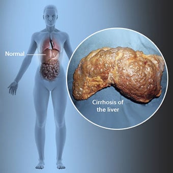
 A cirrhosis a májbetegség szövődménye, amely a májsejtek elvesztésével és a máj visszafordíthatatlan hegesedésével jár.
 A cirrhosisban szenvedő egyéneknél előfordulhat, hogy kevés vagy egyáltalán nem jelentkeznek májbetegség tünetei és jelei. Egyes tünetek nem specifikusak lehetnek, vagyis nem utalnak arra, hogy a máj az oka. A májzsugorodás gyakoribb tünetei és jelei a következők:
A cirrhosisban szenvedő egyéneknél a cirrhosis szövődményeiből származó tünetek és jelek is jelentkeznek.
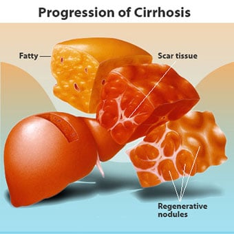 A májzsugorodásnak számos oka van, beleértve a vegyi anyagokat (például alkoholt, zsírt és bizonyos gyógyszereket), vírusokat és mérgező anyagokat. fémek, és autoimmun májbetegség, amelyben a szervezet immunrendszere megtámadja a májat.
 A cirrhosis számos májbetegség szövődménye, amelyet a máj rendellenes szerkezete és működése jellemez. A cirrhosishoz vezető betegségek azért vannak, mert károsítják és elpusztítják a májsejteket, majd a haldokló májsejtekkel kapcsolatos gyulladás és helyreállítás hegszövetek kialakulását idézi elő. Azok a májsejtek, amelyek nem pusztulnak el, szaporodnak, hogy megpróbálják pótolni az elhalt sejteket. Ennek eredményeként újonnan képződött májsejtek (regeneratív csomók) csoportosulnak a hegszövetben. A májzsugornak számos oka van, köztük vegyszerek (például alkohol, zsír és bizonyos gyógyszerek), vírusok, mérgező fémek (például vas és réz, amelyek genetikai betegségek következtében felhalmozódnak a májban), valamint autoimmun májbetegség, amelyben a a szervezet immunrendszere megtámadja a májat.
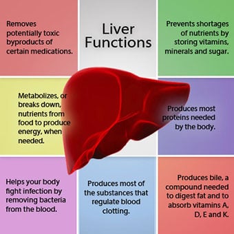 A máj és a vér kapcsolata egyedülálló.
 A máj fontos szerv a szervezetben. Számos kritikus funkciót lát el, amelyek közül kettő a szervezet számára szükséges anyagok előállítása, például a véralvadáshoz szükséges fehérjék véralvadása, valamint a szervezetre ártalmas mérgező anyagok, például gyógyszerek eltávolítása. . A máj fontos szerepet játszik a glükóz (cukor) és lipidek (zsír) ellátásának szabályozásában is, amelyeket a szervezet üzemanyagként használ fel. E kritikus funkciók ellátásához a májsejteknek normálisan kell működniük, és közel kell lenniük a vérhez, mert a máj által hozzáadott vagy eltávolított anyagokat a vér szállítja a májba és onnan. P>
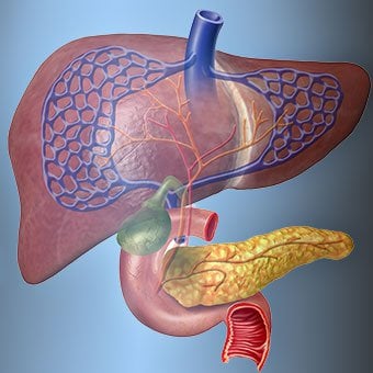
A máj és a vér kapcsolata egyedülálló. A test legtöbb szervétől eltérően az artériák csak kis mennyiségű vért juttatnak a májba. A máj vérellátásának nagy része a bélvénákból származik, amikor a vér visszatér a szívbe. A fő vénát, amely a vért visszavezeti a belekből, portális vénának nevezik. Ahogy a portális véna áthalad a májon, egyre kisebb és kisebb vénákra bomlik. A legapróbb vénák (egyedi felépítésük miatt szinuszoidoknak nevezik) szoros kapcsolatban állnak a májsejtekkel. A májsejtek a szinuszoidok hosszában sorakoznak fel. Ez a szoros kapcsolat a májsejtek és a portális vénából származó vér között lehetővé teszi a májsejtek számára, hogy anyagokat eltávolítsanak és hozzáadjanak a vérhez. Miután a vér áthaladt a szinuszokon, egyre nagyobb és nagyobb vénákba gyűlik össze, amelyek végül egyetlen vénát alkotnak, a májvénát, amely visszavezeti a vért a szívbe.
Cirrózisban a vér és a májsejtek közötti kapcsolat megsemmisül. Annak ellenére, hogy a túlélő vagy újonnan képződött májsejtek képesek lehetnek anyagokat termelni és eltávolítani a vérből, nincs normális, bensőséges kapcsolatuk a vérrel, és ez megzavarja a májsejtek anyagok hozzáadásának vagy eltávolításának képességét. a vértől. Ezenkívül a cirrózisos májban lévő hegesedés akadályozza a vér áramlását a májon és a májsejtek felé. A májon keresztüli véráramlás akadályozása következtében a vér „visszaáll” a portális vénába, és megnő a nyomás a portális vénában, ezt az állapotot portális hipertóniának nevezik. Az áramlás akadályozása és a portális vénában fennálló magas nyomás miatt a portális vénában lévő vér más vénákat keres, amelyekben visszatérhet a szívbe, amelyek alacsonyabb nyomású vénák, amelyek megkerülik a májat. Sajnos a máj nem képes olyan anyagokat hozzáadni vagy eltávolítani a vérből, amelyek megkerülik azt. Ez a májsejtek számának csökkenése, a májon áthaladó vér és a májsejtek közötti normális érintkezés elvesztése, valamint a májat megkerülő vér kombinációja, amely a cirrhosis számos tünetéhez vezet.
A cirrhosis okozta problémák másik oka a májsejtek és az epeáramlási csatornák közötti kapcsolat megzavarása. Az epe a májsejtek által termelt folyadék, amelynek két fontos funkciója van:segíti az emésztést, valamint eltávolítja és eltávolítja a mérgező anyagokat a szervezetből. A májsejtek által termelt epe nagyon apró csatornákba választódik ki, amelyek a szinuszoidokat bélelő májsejtek között futnak, úgynevezett canaliculusokba. A csatornák kis csatornákká ürülnek ki, amelyek azután egyesülnek, és egyre nagyobb csatornákat alkotnak. Az összes csatorna egyetlen csatornává egyesül, amely belép a vékonybélbe, ahol segítheti az élelmiszerek emésztését. Ugyanakkor az epében lévő mérgező anyagok bejutnak a bélbe, majd a széklettel távoznak. Cirrózisban a csatornák rendellenesek, és a májsejtek és a csatornák közötti kapcsolat megsemmisül, akárcsak a májsejtek és a vér kapcsolata a szinuszoidokban. Emiatt a máj nem képes normálisan eltávolítani a mérgező anyagokat, ezek felhalmozódhatnak a szervezetben. Kis mértékben az emésztés a bélben is csökken.
 A májzsugorodás gyakori tünetei és jelei a sárgaság, fáradtság, gyengeség, étvágytalanság, viszketés és könnyű zúzódások.
 A cirrózisban szenvedő betegeknél előfordulhat, hogy a májbetegség tünetei és jelei kevés vagy egyáltalán nem jelentkeznek. Egyes tünetek nem specifikusak lehetnek, és nem utalnak arra, hogy a máj okozza őket. A cirrhosis gyakori tünetei és jelei a következők:
A májzsugorodásban szenvedő betegeknél a betegség szövődményeiből származó tünetek és jelek is jelentkeznek.
A cirrhosis önmagában már a májkárosodás késői szakasza. A májbetegség korai szakaszában májgyulladás lép fel. Ha ezt a gyulladást nem kezelik, hegesedéshez (fibrózishoz) vezethet. Ebben a szakaszban még lehetséges a máj gyógyulása kezeléssel.
Ha a májfibrózist nem kezelik, az cirrózishoz vezethet. Ebben a szakaszban a hegszövet nem tud begyógyulni, de a hegesedés előrehaladása megelőzhető vagy lassítható. Azoknál a cirrhosisban szenvedőknél, akiknél szövődmények jelei mutatkoznak, végstádiumú májbetegség (ESLD) alakulhat ki, és ebben a szakaszban az egyetlen kezelés a májátültetés.
 Ödéma, ascites és bakteriális hashártyagyulladás szövődményei
 Amint a májcirrózis súlyossá válik, a vesék jelzéseket küldenek a só és a víz visszatartására a szervezetben. A felesleges só és víz először a boka és a lábak bőre alatti szövetben halmozódik fel a gravitáció hatására, amikor áll vagy ül. Ezt a folyadék-felhalmozódást perifériás ödémának vagy pitting ödémának nevezik. (A kiütéses ödéma arra utal, hogy az ujjbeggyel erősen megnyomva az ödémás bokát vagy lábszárat bemélyedés keletkezik a bőrön, amely a nyomás feloldása után még egy ideig fennmarad. Bármilyen típusú nyomás, például a zokni rugalmas szalagjából , elegendő lehet a gödrösödéshez.) A duzzanat gyakran rosszabbodik a nap végén, miután felállt vagy ült, és lefekvéskor egyik napról a másikra csökkenhet. A cirrhosis súlyosbodásával és több só és víz visszatartásával a hasfal és a hasi szervek között folyadék is felhalmozódhat (úgynevezett ascites), ami a has duzzanatát, hasi kényelmetlenséget és súlynövekedést okoz.
A hasüregben lévő folyadék (ascites) tökéletes hely a baktériumok szaporodásához. Normális esetben a hasüreg nagyon kis mennyiségű folyadékot tartalmaz, amely jól ellenáll a fertőzéseknek, és a hasüregbe (általában a bélből) bekerülő baktériumok elpusztulnak, vagy eljutnak a portális vénába és a májba, ahol elpusztulnak. Cirrózisban a hasüregben összegyűlő folyadék nem képes normálisan ellenállni a fertőzésnek. Ezenkívül több baktérium jut el a bélből az ascitesbe. A hasüregben és az ascitesben spontán bakteriális peritonitisnek vagy SBP-nek nevezett fertőzés valószínűsíthető. Az SBP életveszélyes szövődmény. Néhány SBP-ben szenvedő betegnek nincsenek tünetei, míg másoknak lázuk, hidegrázásuk, hasi fájdalmuk és érzékenységük, hasmenésük és súlyosbodó ascitesük van.
 Vérzés és lépszövődmények
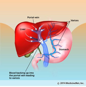
 A cirrózisos májban a hegszövet blokkolja a belekből a szívbe visszatérő vér áramlását, és megemeli a nyomást a portális vénában (portális hipertónia). Ha a nyomás a portális vénában elég magas lesz, akkor a vér a máj körül áramlik, alacsonyabb nyomású vénákon keresztül, hogy elérje a szívet. A leggyakoribb vénák, amelyeken keresztül a vér megkerüli a májat, a nyelőcső alsó részét és a gyomor felső részét bélelő vénák.
A megnövekedett véráramlás és az ebből eredő nyomásnövekedés következtében az alsó nyelőcső és a gyomor felső részének vénái kitágulnak, majd nyelőcső- és gyomorvarixnak nevezik; minél nagyobb a portális nyomás, annál nagyobbak a varixok, és annál valószínűbb, hogy a beteg a varixokból a nyelőcsőbe vagy a gyomorba vérzik.
A varix okozta vérzés súlyos és azonnali kezelés nélkül végzetes lehet. A varixból származó vérzés tünetei közé tartozik a vérhányás (vérrögökkel vagy "kávézaccsal" keveredő vörös vérnek tűnhet), fekete és kátrányos széklet a bélen áthaladó vér változásai miatt (melena), valamint ortosztatikus széklet. szédülés vagy ájulás (a vérnyomás csökkenése okozza, különösen fekvő helyzetből való felálláskor).
Ritkán vérzés léphet fel a belekben máshol képződő varixokból, például a vastagbélben. Az aktívan vérző nyelőcsővarix miatt kórházba került betegeknél nagy a spontán bakteriális hashártyagyulladás kialakulásának kockázata, bár ennek okai még nem tisztázottak.
A lép általában szűrőként működik az idősebb vörösvértestek, fehérvérsejtek és vérlemezkék (a véralvadáshoz fontos apró részecskék) eltávolítására. A lépből kifolyó vér a belekből a portális vénában csatlakozik a vérhez. Ahogy a cirrhosisban megemelkedik a nyomás a portális vénában, egyre inkább gátolja a vér áramlását a lépből. A vér „visszaáll”, felhalmozódik a lépben, és a lép megduzzad, ezt az állapotot lépmegnagyobbodásnak nevezik. Néha a lép annyira megnagyobbodik, hogy hasi fájdalmat okoz.
Ahogy a lép megnagyobbodik, egyre többet szűr ki a vérsejtekből és vérlemezkékből, amíg a vérben le nem csökken a számuk. A hipersplenizmus a kifejezés ennek az állapotnak a leírására, és alacsony vörösvérsejtszámmal (anémia), alacsony fehérvérsejtszámmal (leukopénia) és/vagy alacsony vérlemezkeszámmal (thrombocytopenia) társul. A vérszegénység gyengeséget, a leukopénia fertőzéseket, a thrombocytopenia pedig a véralvadást és elhúzódó vérzést okozhat.
 Máj- (máj) szövődmények
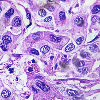
 A bármilyen okból kifolyólag kialakuló cirrhosis növeli az elsődleges májrák (hepatocelluláris karcinóma) kockázatát. Az elsődleges arra a tényre utal, hogy a daganat a májból származik. A másodlagos májrák az, amely a szervezet más részéből származik, és a májba terjed (áttétet képez).
Az elsődleges májrák leggyakoribb tünetei és jelei a hasi fájdalom és duzzanat, a máj megnagyobbodása, a fogyás és a láz. Ezenkívül a májrák számos anyagot termelhet és bocsáthat ki, köztük olyanokat, amelyek megnövekedett vörösvértestszámot (eritrocitózist), alacsony vércukorszintet (hipoglikémia) és magas vér kalciumszintet (hiperkalcémiát) okoznak.
Az élelmiszerben lévő fehérjék egy részét, amely elkerüli az emésztést és a felszívódást, olyan baktériumok használják fel, amelyek általában jelen vannak a bélben. Miközben a fehérjét saját célra használják fel, a baktériumok olyan anyagokat termelnek, amelyeket a bélbe juttatnak, hogy aztán felszívódjanak a szervezetben. Ezen anyagok némelyike, például az ammónia, mérgező hatást gyakorolhat az agyra. Általában ezek a mérgező anyagok a bélből a portális vénán keresztül a májba kerülnek, ahol eltávolítják a vérből és méregtelenítik őket.
Ha cirrózis áll fenn, a májsejtek nem tudnak normálisan működni, vagy azért, mert sérültek, vagy azért, mert elvesztették normális kapcsolatukat a vérrel. Ezenkívül a portális vénában lévő vér egy része megkerüli a májat más vénákon keresztül. Ezeknek a rendellenességeknek az eredménye, hogy a mérgező anyagokat a májsejtek nem tudják eltávolítani, hanem felhalmozódnak a vérben.
Ha a mérgező anyagok kellő mértékben felhalmozódnak a vérben, az agy működése károsodik, ezt az állapotot hepatikus encephalopathiának nevezik. A hepatikus encephalopathia korai tünete a nappali alvás, mint az éjszakai alvás (a normál alvási minta megfordulása). Egyéb tünetek közé tartozik az ingerlékenység, a koncentrálóképesség vagy a számítások végzésére való képtelenség, a memóriavesztés, a zavartság vagy a depressziós tudatszint. Végül a súlyos hepatikus encephalopathia kómát és halált okoz.
A mérgező anyagok a májzsugorodásban szenvedő betegek agyát is nagyon érzékennyé teszik azokra a gyógyszerekre, amelyeket általában a máj szűr és méregtelenít. Lehetséges, hogy számos gyógyszer adagját csökkenteni kell, hogy elkerüljük a mérgező cirrhosis felhalmozódását, különösen a nyugtatók és az elalvást elősegítő gyógyszerek. Alternatív megoldásként olyan gyógyszerek is használhatók, amelyeket nem kell méregteleníteni vagy a májnak kiüríteni a szervezetből, ilyenek például a vesék által kiürülő gyógyszerek.
A súlyosbodó cirrhosisban szenvedő betegeknél hepatorenalis szindróma alakulhat ki. Ez a szindróma súlyos szövődmény, amelyben a vesék működése csökken. Ez egy funkcionális probléma a vesékben, vagyis nincs fizikai károsodás a vesében. Ehelyett a csökkent funkció a veséken keresztüli véráramlás megváltozásának köszönhető. A hepatorenalis szindrómát úgy határozzák meg, hogy a vesék progresszív kudarca az anyagok vérből való eltávolításában és megfelelő mennyiségű vizelet előállításában, miközben a vese egyéb fontos funkciói, például a sóvisszatartás megmarad. Ha a májfunkció javul vagy egészséges májat ültetnek át egy hepatorenalis szindrómában szenvedő betegbe, a vesék általában újra normálisan működnek. Ez arra utal, hogy a vesék csökkent működése vagy a mérgező anyagok felhalmozódása a vérben, vagy a májelégtelenség miatti kóros májműködés eredménye. A hepatorenalis szindrómának két típusa van. Az egyik típus fokozatosan, hónapok alatt jelentkezik. A másik gyorsan egy-két hét alatt következik be.
Ritkán egyes előrehaladott cirrhosisban szenvedő betegeknél hepatopulmonalis szindróma alakulhat ki. Ezek a betegek légzési nehézséget tapasztalhatnak, mivel bizonyos, előrehaladott cirrhosisban felszabaduló hormonok a tüdő rendellenes működését okozzák. A tüdőben az alapvető probléma, hogy a tüdőben a tüdő alveolusaival (légzsákokkal) érintkező kis vérereken keresztül nem áramlik elég vér. A tüdőn átáramló vér az alveolusok körül söntölődik, és nem tud elegendő oxigént felvenni az alveolusokban lévő levegőből. Ennek eredményeként a beteg légszomjat tapasztal, különösen terheléskor.
 A cirrózisnak 12 gyakori oka van.
 Common causes of cirrhosis of the liver include:
Less common causes of cirrhosis include:
In certain parts of the world (particularly Northern Africa), infection of the liver with a parasite (schistosomiasis) is the most common cause of liver disease and cirrhosis.
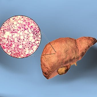 Alcohol and nonalcoholic fatty liver disease are common causes of cirrhosis.
 Alcohol is a very common cause of cirrhosis, particularly in the Western world. Chronic, high levels of alcohol consumption injure liver cells. Thirty percent of individuals who drink daily at least eight to sixteen ounces of hard liquor or the equivalent for fifteen or more years will develop cirrhosis. Alcohol causes a range of liver diseases, which include simple and uncomplicated fatty liver (steatosis), more serious fatty liver with inflammation (steatohepatitis or alcoholic hepatitis), and cirrhosis.
Nonalcoholic fatty liver disease (NAFLD) refers to a wide spectrum of liver diseases that, like alcoholic liver disease, range from simple steatosis, to nonalcoholic steatohepatitis (NASH), to cirrhosis. All stages of NAFLD have in common the accumulation of fat in liver cells. The term nonalcoholic is used because NAFLD occurs in individuals who do not consume excessive amounts of alcohol, yet in many respects the microscopic picture of NAFLD is similar to what can be seen in liver disease that is due to excessive alcohol. NAFLD is associated with a condition called insulin resistance, which, in turn, is associated with metabolic syndrome and diabetes mellitus type 2. Obesity is the main cause of insulin resistance, metabolic syndrome, and type 2 diabetes. NAFLD is the most common liver disease in the United States and is responsible for up to 25% of all liver disease. The number of livers transplanted for NAFLD-related cirrhosis is on the rise. Public health officials are worried that the current epidemic of obesity will dramatically increase the development of NAFLD and cirrhosis in the population.
 Primary biliary cirrhosis (PBC) is a liver disease caused by an abnormality of the immune system that is found predominantly in women.
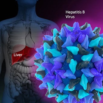
 Chronic viral hepatitis is a condition in which hepatitis B or hepatitis C virus infects the liver for years. Most patients with viral hepatitis will not develop chronic hepatitis and cirrhosis. The majority of patients infected with hepatitis A recover completely within weeks, without developing chronic infection. In contrast, some patients infected with hepatitis B virus and most patients infected with hepatitis C virus develop chronic hepatitis, which, in turn, causes progressive liver damage and leads to cirrhosis, and, sometimes, liver cancers.
Autoimmune hepatitis is a liver disease found more commonly in women that is caused by an abnormality of the immune system. The abnormal immune activity in autoimmune hepatitis causes progressive inflammation and destruction of liver cells (hepatocytes), leading ultimately to cirrhosis.
Primary biliary cirrhosis (PBC) is a liver disease caused by an abnormality of the immune system that is found predominantly in women. The abnormal immunity in PBC causes chronic inflammation and destruction of the small bile ducts within the liver. The bile ducts are passages within the liver through which bile travels to the intestine. Bile is a fluid produced by the liver that contains substances required for digestion and absorption of fat in the intestine, as well as other compounds that are waste products, such as the pigment bilirubin. (Bilirubin is produced by the breakdown of hemoglobin from old red blood cells.). Along with the gallbladder, the bile ducts make up the biliary tract. In PBC, the destruction of the small bile ducts blocks the normal flow of bile into the intestine. As the inflammation continues to destroy more of the bile ducts, it also spreads to destroy nearby liver cells. As the destruction of the hepatocytes proceeds, scar tissue (fibrosis) forms and spreads throughout the areas of destruction. The combined effects of progressive inflammation, scarring, and the toxic effects of accumulating waste products culminates in cirrhosis.
Primary sclerosing cholangitis (PSC) is an uncommon disease frequently found in patients with Crohn's disease and ulcerative colitis. In PSC, the large bile ducts outside of the liver become inflamed, narrowed, and obstructed. Obstruction to the flow of bile leads to infections of the bile ducts and jaundice, eventually causing cirrhosis. In some patients, injury to the bile ducts (usually because of surgery) also can cause obstruction and cirrhosis of the liver.
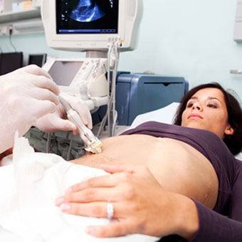 Different liver diseases should be diagnosed by specialists and different tests such as liver blood test, biopsy, and others.
 Inherited (genetic) disorders that result in the accumulation of toxic substances in the liver, which leads to tissue damage and cirrhosis. Examples include the abnormal accumulation of iron (hemochromatosis) or copper (Wilson disease). In hemochromatosis, patients inherit a tendency to absorb an excessive amount of iron from food. Over time, iron accumulation in different organs throughout the body causes cirrhosis, arthritis, heart muscle damage leading to heart failure, and testicular dysfunction causing loss of sexual drive. Treatment is aimed at preventing damage to organs by removing iron from the body through phlebotomy (removing blood). In Wilson disease, there is an inherited abnormality in one of the proteins that control copper in the body. Over time, copper accumulates in the liver, eyes, and brain. Cirrhosis, tremor, psychiatric disturbances, and other neurological difficulties occur if the condition is not treated early. Treatment is with oral medication, which increases the amount of copper that is eliminated from the body in the urine.
Cryptogenic cirrhosis (cirrhosis due to unidentified causes) is a common reason for liver transplantation. It is termed called cryptogenic cirrhosis because for many years doctors have been being unable to explain why a proportion of patients developed cirrhosis. Doctors now believe that cryptogenic cirrhosis is due to NASH (nonalcoholic steatohepatitis) caused by long-standing obesity, type 2 diabetes, and insulin resistance. The fat in the liver of patients with NASH is believed to disappear with the onset of cirrhosis, and this has made it difficult for doctors to make the connection between NASH and cryptogenic cirrhosis for a long time. One important clue that NASH leads to cryptogenic cirrhosis is the finding of a high occurrence of NASH in the new livers of patients undergoing liver transplant for cryptogenic cirrhosis. Finally, a study from France suggests that patients with NASH have a similar risk of developing cirrhosis as patients with long-standing infection with hepatitis C virus. (See discussion that follows.) However, the progression to cirrhosis from NASH is thought to be slow and the diagnosis of cirrhosis typically is made in people in their sixties.
Infants can be born without bile ducts (biliary atresia) and ultimately develop cirrhosis. Other infants are born lacking vital enzymes for controlling sugars that lead to the accumulation of sugars and cirrhosis. On rare occasions, the absence of a specific enzyme can cause cirrhosis and scarring of the lung (alpha-1 antitrypsin deficiency).
Less common causes of cirrhosis include unusual reactions to some drugs and prolonged exposure to toxins, as well as chronic heart failure (cardiac cirrhosis). In certain parts of the world (particularly Northern Africa), infection of the liver with a parasite (schistosomiasis) is the most common cause of liver disease and cirrhosis.
 Different liver diseases should be diagnosed by specialists and different tests such as liver blood test, biopsy, and others.
 The single best test for diagnosing cirrhosis is a biopsy of the liver. Liver biopsies carry a small risk for serious complications, and biopsy often is reserved for those patients in whom the diagnosis of the type of liver disease or the presence of cirrhosis is not clear. The history, physical examination, or routine testing may suggest the possibility of cirrhosis. If cirrhosis is present, other tests can be used to determine the severity of the cirrhosis and the presence of complications. Tests also may be used to diagnose the underlying disease that is causing the cirrhosis. Examples of how doctors diagnose and evaluate cirrhosis are:
 There are four types of treatment of cirrhosis.
 Treatment of cirrhosis includes
Consume a balanced diet and one multivitamin daily. Patients with PBC with impaired absorption of fat-soluble vitamins may need additional vitamins D and K.
Avoid drugs (including alcohol) that cause liver damage. All patients with cirrhosis should avoid alcohol. Most patients with alcohol-induced cirrhosis experience an improvement in liver function with abstinence from alcohol. Even patients with chronic hepatitis B and C can substantially reduce liver damage and slow the progression towards cirrhosis with abstinence from alcohol.
Avoid nonsteroidal anti-inflammatory drugs (NSAIDs, e.g., ibuprofen). Patients with cirrhosis can experience worsening of liver and kidney function with NSAIDs.
Eradicate hepatitis B and hepatitis C virus by using anti-viral medications. Not all patients with cirrhosis due to chronic viral hepatitis are candidates for drug treatment. Some patients may experience serious deterioration in liver function and/or intolerable side effects during treatment. Thus, decisions to treat viral hepatitis have to be individualized, after consulting with doctors experienced in treating liver diseases (hepatologists).
Remove blood from patients with hemochromatosis to reduce the levels of iron and prevent further damage to the liver. In Wilson's disease, medications can be used to increase the excretion of copper in the urine to reduce the levels of copper in the body and prevent further damage to the liver.
Suppress the immune system with drugs such as prednisone and azathioprine (Imuran) to decrease inflammation of the liver in autoimmune hepatitis.
Treat patients with PBC with a bile acid preparation, ursodeoxycholic acid (UDCA), also called ursodiol (Actigall). Results of an analysis that combined the results from several clinical trials showed that UDCA increased survival among PBC patients during 4 years of therapy. The development of portal hypertension also was reduced by the UDCA. It is important to note that despite producing clear benefits, UDCA treatment primarily retards progression and does not cure PBC. Other medications such as colchicine and methotrexate also may have benefits in subsets of patients with PBC.
Immunize patients with cirrhosis against infection with hepatitis A and B to prevent a serious deterioration in liver. There are currently no vaccines available for immunizing against hepatitis C.
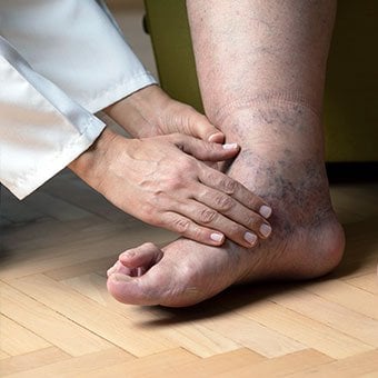 Treatment for edema, ascites, and hypersplenism complications.
 Retaining salt and water can lead to swelling of the ankles and legs (edema) or abdomen (ascites) in patients with cirrhosis. Doctors often advise patients with cirrhosis to restrict dietary salt (sodium) and fluid to decrease edema and ascites. The amount of salt in the diet usually is restricted to 2 grams per day and fluid to 1.2 liters per day. In most patients with cirrhosis, salt and fluid restriction is not enough and diuretics have to be added.
Diuretics are medications that work in the kidneys to promote the elimination of salt and water into the urine. A combination of the diuretics spironolactone (Aldactone) and furosemide (Lasix) can reduce or eliminate the edema and ascites in most patients. During treatment with diuretics, it is important to monitor the function of the kidneys by measuring blood levels of blood urea nitrogen (BUN) and creatinine to determine if too much diuretic is being used. Too much diuretic can lead to kidney dysfunction that is reflected in elevations of the BUN and creatinine levels in the blood.
Sometimes, when the diuretics do not work (in which case the ascites is said to be refractory), a long needle or catheter is used to draw out the ascitic fluid directly from the abdomen, a procedure called abdominal paracentesis. It is common to withdraw large amounts (liters) of fluid from the abdomen when the ascites is causing painful abdominal distension and/or difficulty breathing because it limits the movement of the diaphragms.
Another treatment for refractory ascites is a procedure called transjugular intravenous portosystemic shunting (TIPS).
The spleen normally acts as a filter to remove older red blood cells, white blood cells, and platelets (small particles important for the clotting of blood). The blood that drains from the spleen joins the blood in the portal vein from the intestines. As the pressure in the portal vein rises in cirrhosis, it increasingly blocks the flow of blood from the spleen. The blood "backs-up," accumulating in the spleen, and the spleen swells in size, a condition referred to as splenomegaly. Sometimes, the spleen is so enlarged it causes abdominal pain.
As the spleen enlarges, it filters out more and more of the blood cells and platelets until their numbers in the blood are reduced. Hypersplenism is the term used to describe this condition, and it is associated with a low red blood cell count (anemia), low white blood cell count (leukopenia), and/or a low platelet count (thrombocytopenia). Anemia can cause weakness, leucopenia can lead to infections, and thrombocytopenia can impair the clotting of blood and result in prolonged bleeding.
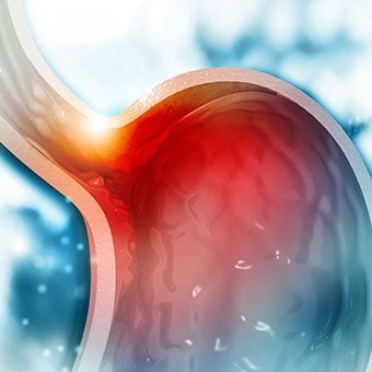 Once varices have bled, they tend to rebleed and the probability that a patient will die from each bleeding episode is high (30% to 35%). Treatment is necessary to prevent the first bleeding episode as well as rebleeding.
 If large varices develop in the esophagus or upper stomach, patients with cirrhosis are at risk for serious bleeding due to rupture of these varices. Once varices have bled, they tend to rebleed and the probability that a patient will die from each bleeding episode is high (30% to 35%). Treatment is necessary to prevent the first bleeding episode as well as rebleeding. Treatments include medications and procedures to decrease the pressure in the portal vein and procedures to destroy the varices.
 Hepatic encephalopathy usually should be treated with a low protein diet and oral lactulose.
 Patients with an abnormal sleep cycle, impaired thinking, odd behavior, or other signs of hepatic encephalopathy usually should be treated with a low protein diet and oral lactulose. Dietary protein is restricted because it is a source of toxic compounds that cause hepatic encephalopathy. Lactulose, which is a liquid, traps toxic compounds in the colon so they cannot be absorbed into the bloodstream, and thus cause encephalopathy. Lactulose is converted to lactic acid in the colon, and the acidic environment that results is believed to trap the toxic compounds produced by the bacteria. To be sure adequate lactulose is present in the colon at all times, the patient should adjust the dose to produce 2 to 3 semiformed bowel movements a day. Lactulose is a laxative, and the effectiveness of treatment can be judged by loosening or increasing the frequency of stools. Rifaximin (Xifaxan) is an antibiotic taken orally that is not absorbed into the body but rather remains in the intestines. It is the preferred mode of treatment of hepatic encephalopathy. Antibiotics work by suppressing the bacteria that produce the toxic compounds in the colon.
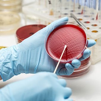 Most patients with spontaneous bacterial peritonitis are hospitalized and treated with intravenous antibiotics.
 Patients suspected of having spontaneous bacterial peritonitis usually will undergo paracentesis. The fluid that is removed is examined for white blood cells and cultured for bacteria. Culturing involves inoculating a sample of the ascites into a bottle of nutrient-rich fluid that encourages the growth of bacteria, thus facilitating the identification of even small numbers of bacteria. Blood and urine samples also are often obtained for culturing because many patients with spontaneous bacterial peritonitis also will have infections in their blood and urine. Many doctors believe the infection may have begun in the blood and the urine and spread to the ascitic fluid to cause spontaneous bacterial peritonitis. Most patients with spontaneous bacterial peritonitis are hospitalized and treated with intravenous antibiotics such as cefotaxime (Claforan). Patients usually treated with antibiotics include:
Spontaneous bacterial peritonitis is a serious infection. It often occurs in patients with advanced cirrhosis whose immune systems are weak, but with modern antibiotics and early detection and treatment, the prognosis of recovering from an episode of spontaneous bacterial peritonitis is good.
In some patients, oral antibiotics (norfloxacin [Noroxin] or sulfamethoxazole and trimethoprim [Bactrim]) can be prescribed to prevent spontaneous bacterial peritonitis. Not all patients with cirrhosis and ascites should be treated with antibiotics to prevent spontaneous bacterial peritonitis, but some patients are at high risk for developing spontaneous bacterial peritonitis and warrant preventive treatment.
 The prognosis and life expectancy for cirrhosis of the liver varies and depends on the cause, the severity, any complications, and any underlying diseases.
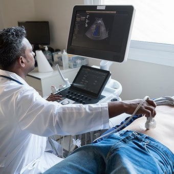
 Several types of liver disease that cause cirrhosis (such as hepatitis B and C) are associated with a high incidence of liver cancer. It is useful to screen for liver cancer in patients with cirrhosis, as early surgical treatment or transplantation of the liver can cure the patient of cancer. The difficulty is that the methods available for screening are only partially effective, identifying at best only half of patients at a curable stage of their cancer. Despite the partial effectiveness of screening, most patients with cirrhosis, particularly hepatitis B and C, are screened yearly or every six months with ultrasound examination of the liver and measurements of cancer-produced proteins in the blood, for example, alpha-fetoprotein.
Cirrhosis is irreversible. Liver function usually gradually worsens despite treatment, and complications of cirrhosis increase and become difficult to treat. When cirrhosis is far advanced liver transplantation often is the only option for treatment. Recent advances in surgical transplantation and medications to prevent infection and rejection of the transplanted liver have greatly improved survival after transplantation. On average, more than 80% of patients who receive transplants are alive after five years. Not everyone with cirrhosis is a candidate for transplantation. Furthermore, there is a shortage of livers to transplant, and they're usually is a long (months to years) wait before a liver for transplanting becomes available. Measures to slow the progression of liver disease, and treat and prevent complications of cirrhosis are vitally important.
The prognosis and life expectancy for cirrhosis of the liver varies and depends on the cause, the severity, any complications, and any underlying diseases.
Progress in the management and prevention of cirrhosis continues. Research is ongoing to determine the mechanism of scar formation in the liver and how this process of scarring can be interrupted or even reversed. Newer and better treatments for viral liver disease are being developed to prevent the progression to cirrhosis. Prevention of viral hepatitis by vaccination, which is available for hepatitis B, is being developed for hepatitis C. Treatments for the complications of cirrhosis are being developed or revised, and tested continually. Finally, research is being directed at identifying new proteins in the blood that can detect liver cancer early or predict which patients will develop liver cancer.
 Antibiotikumok gabonával táplált húsban:Amit tudnod kell
 Intézkedések a SARS-CoV-2 szennyvízen keresztüli terjedésének megakadályozására a szegény régiókban
 Alacsony FODMAP-tartalmú diéta IBS esetén:Az enni és kerülendő élelmiszerek listája
 A sok vörös hús a férfiak bélrendszeri rendellenességeihez köthető
 A szivárgó bél és a mikrobiális dysbiosis hozzájárulhat a citokin viharhoz súlyosan beteg COVID-19 esetekben
 Mire számíthat a székletátültetés?
 Új eszköz rögzíti és követi a mikrobióma növekedését
Az elmúlt években, az emberi mikrobiom óriási népszerűségre tett szert az egészségének alakításában játszott szerepe miatt. Elengedhetetlen az emberi fejlődéshez, táplálás, és az immunitás. Ezért sok
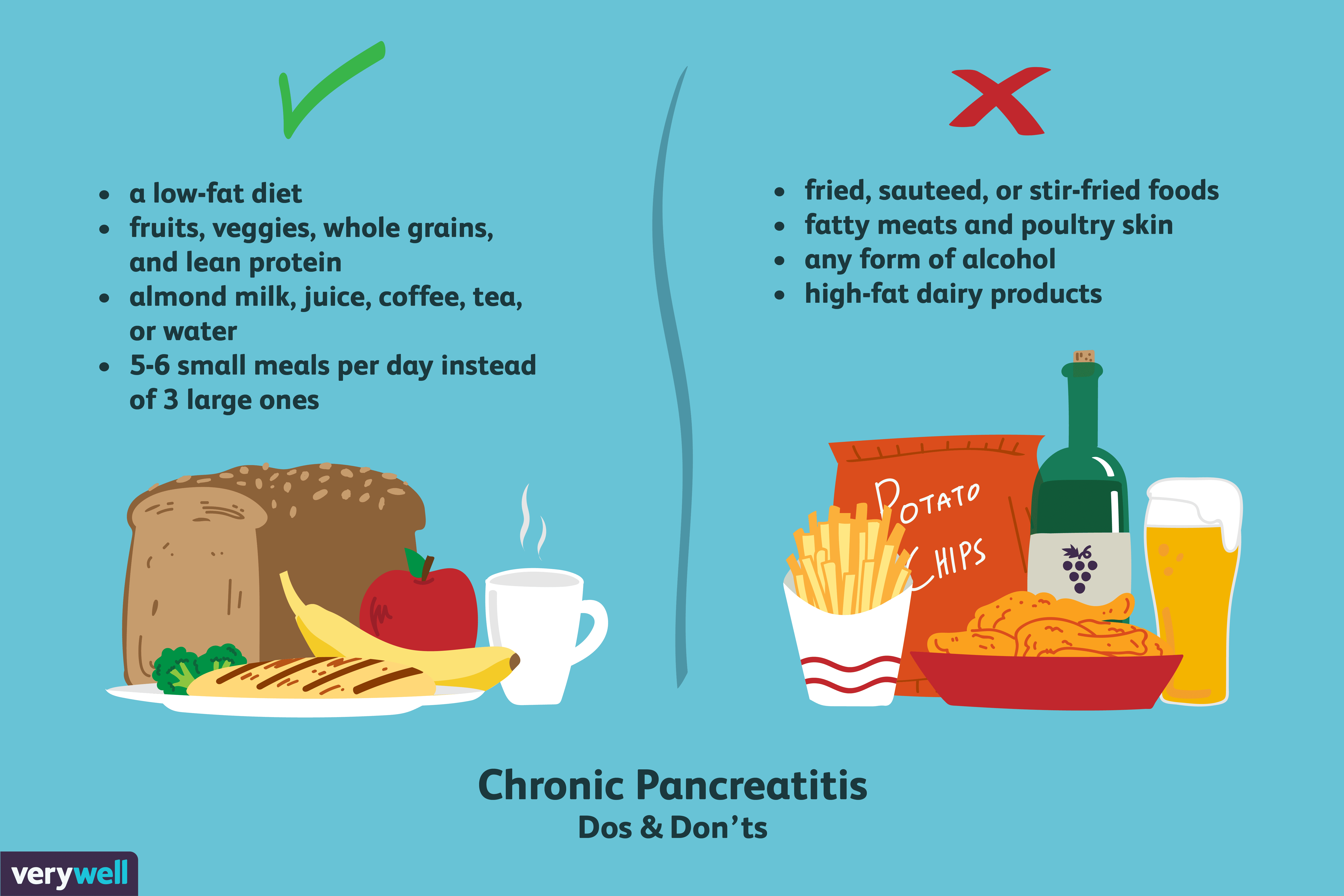 Mit együnk, ha hasnyálmirigy-gyulladása van?
Az inzulin, a szervezet által a vércukorszint szabályozására használt hormon termelése mellett az egészséges hasnyálmirigy enzimeket is termel, amelyek segítik a szervezet emésztését és az elfogyaszto
 1. hét értékelése a 8 hetes vércukor-diétáról
Ez az első hete Dr. Michael Mosleyt programja:A 8 hetes vércukor-diéta . A Vékonybél-baktériumok túlszaporodása (SIBO) miatt felgyorsult súly csökkentése érdekében. , követni fogom a800 kalóriát napon